• 文献检索
  • 文档翻译
  • 深度研究
  • 学术资讯
  • Suppr Zotero 插件Zotero 插件
  • 邀请有礼
  • 套餐&价格
  • 历史记录
应用&插件
Suppr Zotero 插件Zotero 插件浏览器插件Mac 客户端Windows 客户端微信小程序
定价
高级版会员购买积分包购买API积分包
服务
文献检索文档翻译深度研究API 文档MCP 服务
关于我们
关于 Suppr公司介绍联系我们用户协议隐私条款
关注我们

Suppr 超能文献

核心技术专利:CN118964589B侵权必究
粤ICP备2023148730 号-1Suppr @ 2026

文献检索

告别复杂PubMed语法,用中文像聊天一样搜索,搜遍4000万医学文献。AI智能推荐,让科研检索更轻松。

立即免费搜索

文件翻译

保留排版,准确专业,支持PDF/Word/PPT等文件格式,支持 12+语言互译。

免费翻译文档

深度研究

AI帮你快速写综述,25分钟生成高质量综述,智能提取关键信息,辅助科研写作。

立即免费体验

系统蛋白质组学评估小胶质细胞对肿瘤支持性抗炎细胞因子的反应。

Systems-Level Proteomics Evaluation of Microglia Response to Tumor-Supportive Anti-Inflammatory Cytokines.

机构信息

Department of Biological Sciences, Virginia Tech, Blacksburg, VA, United States.

Fralin Life Sciences Institute, Virginia Tech, Blacksburg, VA, United States.

出版信息

Front Immunol. 2021 Sep 9;12:646043. doi: 10.3389/fimmu.2021.646043. eCollection 2021.

DOI:10.3389/fimmu.2021.646043
PMID:34566949
原文链接:https://pmc.ncbi.nlm.nih.gov/articles/PMC8458581/
Abstract

BACKGROUND

Microglia safeguard the CNS against injuries and pathogens, and in the presence of certain harmful stimuli are capable of inducing a disease-dependent inflammatory response. When exposed to anti-inflammatory cytokines, however, these cells possess the ability to switch from an inflammatory to an immunosuppressive phenotype. Cancer cells exploit this property to evade the immune system, and elicit an anti-inflammatory microenvironment that facilitates tumor attachment and growth.

OBJECTIVE

The tumor-supportive biological processes that are activated in microglia cells in response to anti-inflammatory cytokines released from cancer cells were explored with mass spectrometry and proteomic technologies.

METHODS

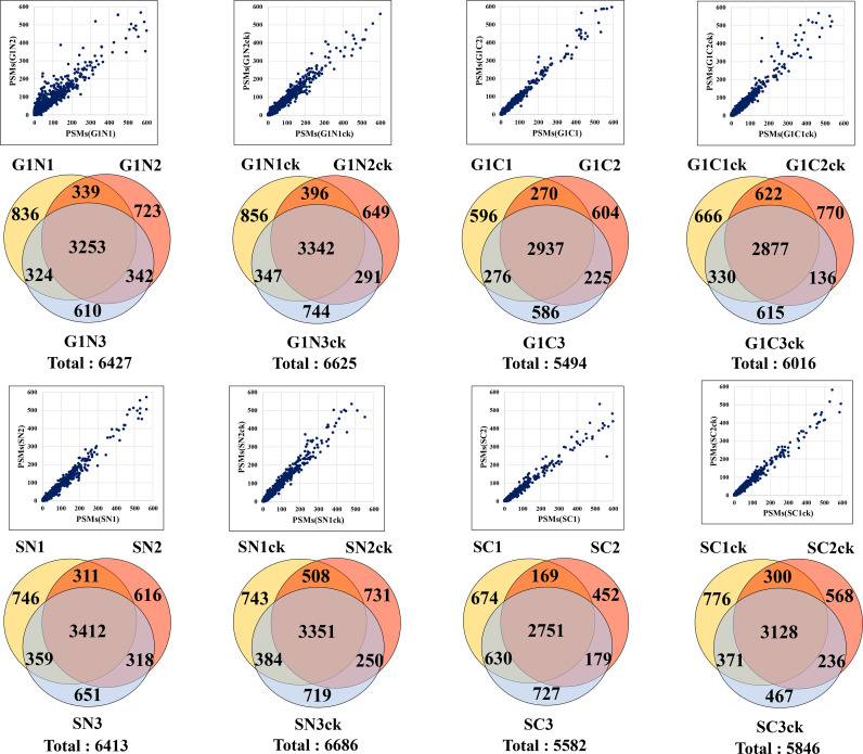
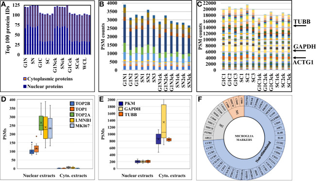
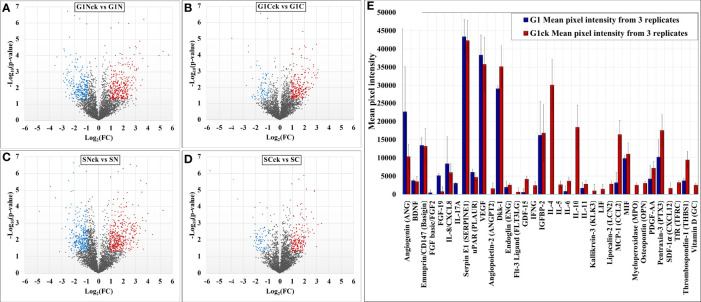
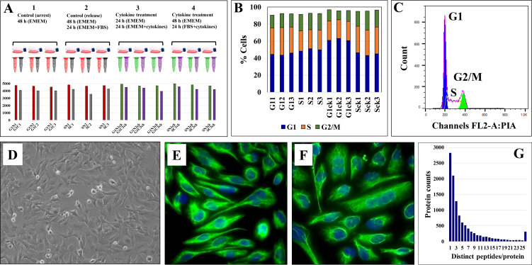
Serum-depleted and non-depleted human microglia cells (HMC3) were treated with a cocktail of IL-4, IL-13, IL-10, TGFβ, and CCL2. The cellular protein extracts were analyzed by LC-MS/MS. Using functional annotation clustering tools, statistically significant proteins that displayed a change in abundance between cytokine-treated and non-treated cells were mapped to their biological networks and pathways.

RESULTS

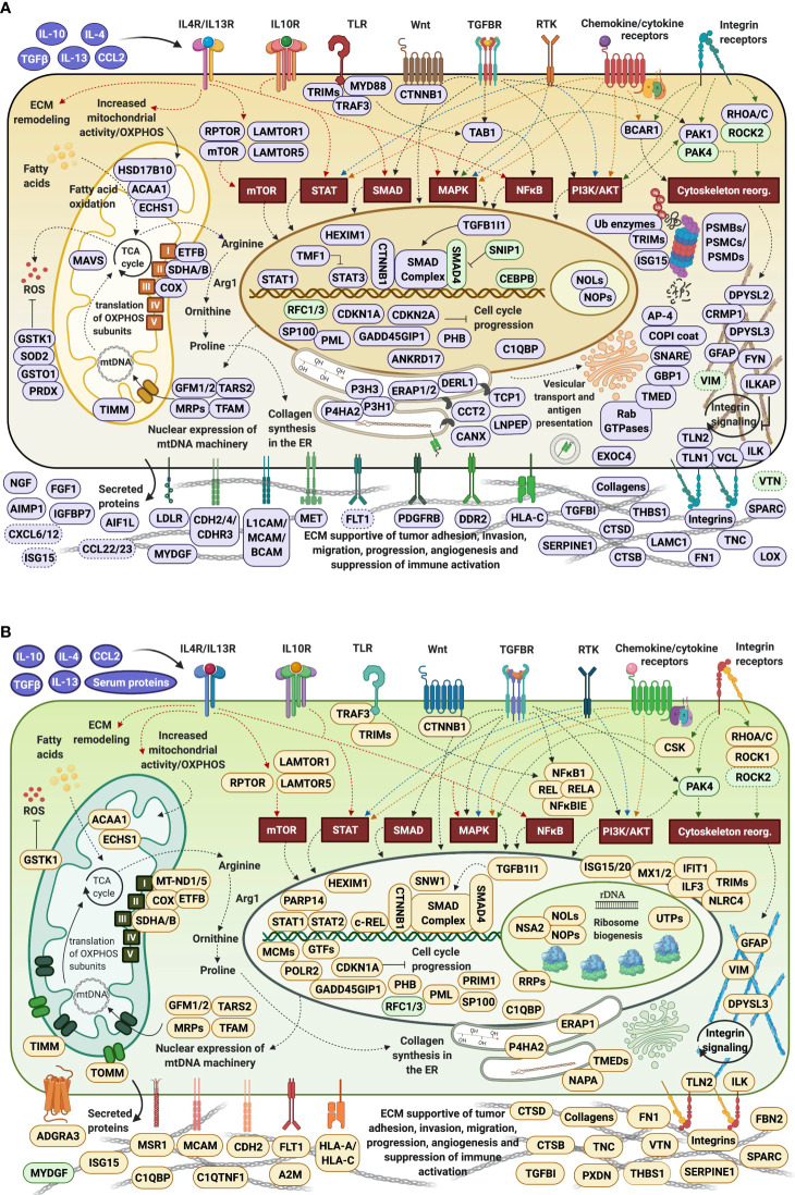
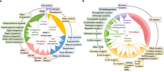
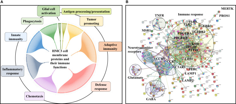
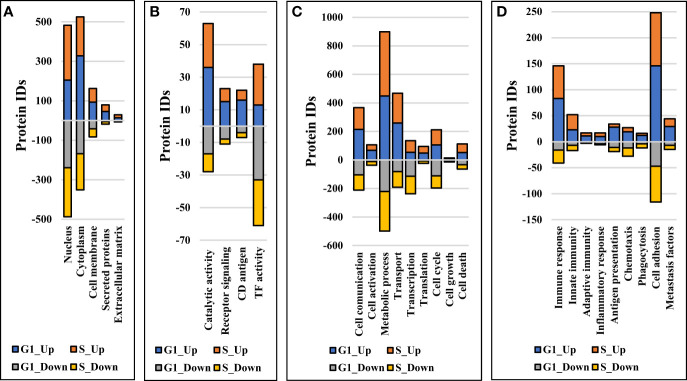
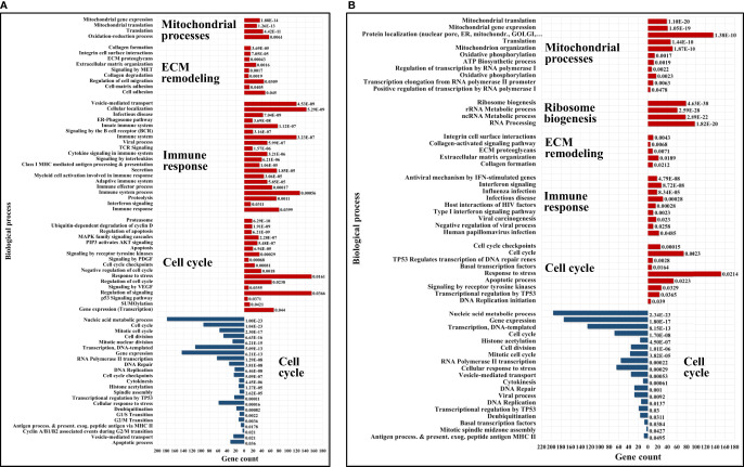
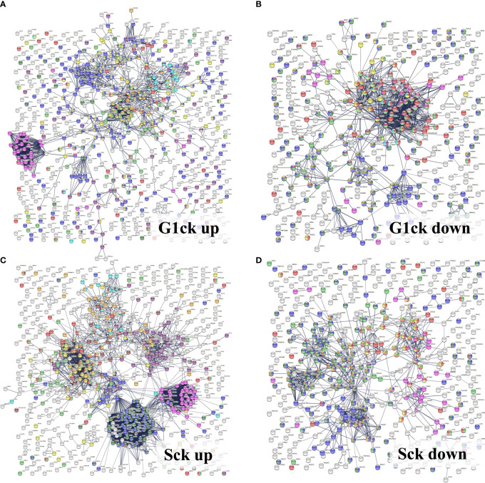
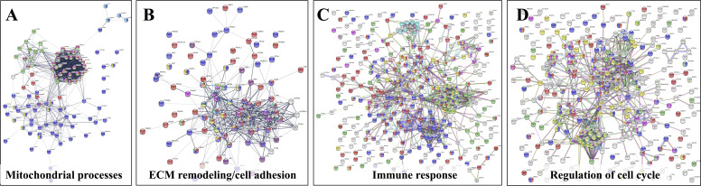
The proteomic analysis of HMC3 cells enabled the identification of ~10,000 proteins. Stimulation with anti-inflammatory cytokines resulted in the activation of distinct, yet integrated clusters of proteins that trigger downstream a number of tumor-promoting biological processes. The observed changes could be classified into four major categories, i.e., mitochondrial gene expression, ECM remodeling, immune response, and impaired cell cycle progression. Intracellular immune activation was mediated mainly by the transducers of MAPK, STAT, TGFβ, NFKB, and integrin signaling pathways. Abundant collagen formation along with the expression of additional receptors, matrix components, growth factors, proteases and protease inhibitors, was indicative of ECM remodeling processes supportive of cell-cell and cell-matrix adhesion. Overexpression of integrins and their modulators was reflective of signaling processes that link ECM reorganization with cytoskeletal re-arrangements supportive of cell migration. Antigen processing/presentation was represented by HLA class I histocompatibility antigens, and correlated with upregulated proteasomal subunits, vesicular/viral transport, and secretory processes. Immunosuppressive and proangiogenic chemokines, as well as anti-angiogenic factors, were detectable in low abundance. Pronounced pro-inflammatory, chemotactic or phagocytic trends were not observed, however, the expression of certain receptors, signaling and ECM proteins indicated the presence of such capabilities.

CONCLUSIONS

Comprehensive proteomic profiling of HMC3 cells stimulated with anti-inflammatory cytokines revealed a spectrum of microglia phenotypes supportive of cancer development in the brain microenvironment-dependent biological mechanisms.

https://cdn.ncbi.nlm.nih.gov/pmc/blobs/df78/8458581/bb8928613930/fimmu-12-646043-g011.jpg
https://cdn.ncbi.nlm.nih.gov/pmc/blobs/df78/8458581/2fc52ecdff38/fimmu-12-646043-g001.jpg
https://cdn.ncbi.nlm.nih.gov/pmc/blobs/df78/8458581/9f6608e876fc/fimmu-12-646043-g002.jpg
https://cdn.ncbi.nlm.nih.gov/pmc/blobs/df78/8458581/91e11088742b/fimmu-12-646043-g003.jpg
https://cdn.ncbi.nlm.nih.gov/pmc/blobs/df78/8458581/8b3bc614c160/fimmu-12-646043-g004.jpg
https://cdn.ncbi.nlm.nih.gov/pmc/blobs/df78/8458581/5f4e3e4cf0fe/fimmu-12-646043-g005.jpg
https://cdn.ncbi.nlm.nih.gov/pmc/blobs/df78/8458581/2520e9b80395/fimmu-12-646043-g006.jpg
https://cdn.ncbi.nlm.nih.gov/pmc/blobs/df78/8458581/71c0b72368a8/fimmu-12-646043-g007.jpg
https://cdn.ncbi.nlm.nih.gov/pmc/blobs/df78/8458581/c216f3416f72/fimmu-12-646043-g008.jpg
https://cdn.ncbi.nlm.nih.gov/pmc/blobs/df78/8458581/f6e123cba151/fimmu-12-646043-g009.jpg
https://cdn.ncbi.nlm.nih.gov/pmc/blobs/df78/8458581/8fa857e90aa0/fimmu-12-646043-g010.jpg
https://cdn.ncbi.nlm.nih.gov/pmc/blobs/df78/8458581/bb8928613930/fimmu-12-646043-g011.jpg
摘要

背景

小胶质细胞保护中枢神经系统免受损伤和病原体的侵害,并且在存在某些有害刺激的情况下,能够诱导依赖于疾病的炎症反应。然而,当暴露于抗炎细胞因子时,这些细胞具有从炎症状态向免疫抑制状态转变的能力。癌细胞利用这种特性来逃避免疫系统,并引发有利于肿瘤附着和生长的抗炎微环境。

目的

利用质谱和蛋白质组学技术探索小胶质细胞在响应癌细胞释放的抗炎细胞因子时激活的支持肿瘤的生物学过程。

方法

用包含 IL-4、IL-13、IL-10、TGFβ 和 CCL2 的细胞因子鸡尾酒处理血清耗尽和非耗尽的人小胶质细胞(HMC3)。通过 LC-MS/MS 分析细胞蛋白提取物。使用功能注释聚类工具,将细胞因子处理和未处理细胞之间丰度变化的具有统计学意义的蛋白质映射到它们的生物网络和途径。

结果

对 HMC3 细胞的蛋白质组学分析能够鉴定出约 10000 种蛋白质。用抗炎细胞因子刺激导致不同但整合的蛋白质簇的激活,触发了许多促进肿瘤的生物学过程。观察到的变化可以分为四大类,即线粒体基因表达、ECM 重塑、免疫反应和细胞周期进程受损。细胞内免疫激活主要由 MAPK、STAT、TGFβ、NFKB 和整合素信号通路的转导子介导。丰富的胶原蛋白形成以及其他受体、基质成分、生长因子、蛋白酶和蛋白酶抑制剂的表达表明 ECM 重塑过程支持细胞-细胞和细胞-基质附着。整合素及其调节剂的过度表达反映了将 ECM 重排与支持细胞迁移的细胞骨架重新排列联系起来的信号过程。抗原加工/呈递由 HLA I 类组织相容性抗原代表,与上调的蛋白酶体亚基、囊泡/病毒运输和分泌过程相关。可检测到低丰度的免疫抑制和促血管生成趋化因子以及抗血管生成因子。然而,某些受体、信号和 ECM 蛋白的表达表明存在这种能力,并未观察到明显的促炎、趋化或吞噬趋势。

结论

用抗炎细胞因子刺激 HMC3 细胞的全面蛋白质组学分析揭示了一系列支持大脑中癌症发展的小胶质细胞表型,其机制依赖于大脑微环境。

相似文献

1
Systems-Level Proteomics Evaluation of Microglia Response to Tumor-Supportive Anti-Inflammatory Cytokines.系统蛋白质组学评估小胶质细胞对肿瘤支持性抗炎细胞因子的反应。
Front Immunol. 2021 Sep 9;12:646043. doi: 10.3389/fimmu.2021.646043. eCollection 2021.
2
Anti-inflammatory cytokine stimulation of HMC3 cells: Proteome dataset.人单核细胞系HMC3细胞的抗炎细胞因子刺激:蛋白质组数据集
Data Brief. 2023 Jul 20;49:109433. doi: 10.1016/j.dib.2023.109433. eCollection 2023 Aug.
3
Interferon regulatory factor 3 plays an anti-inflammatory role in microglia by activating the PI3K/Akt pathway.干扰素调节因子 3 通过激活 PI3K/Akt 通路在小胶质细胞中发挥抗炎作用。
J Neuroinflammation. 2011 Dec 30;8:187. doi: 10.1186/1742-2094-8-187.
4
Profiling the proteomic inflammatory state of human astrocytes using DIA mass spectrometry.采用 DIA 质谱技术对人星形胶质细胞的蛋白质组炎症状态进行分析。
J Neuroinflammation. 2018 Nov 30;15(1):331. doi: 10.1186/s12974-018-1371-6.
5
A systems pharmacology-based approach to identify novel Kv1.3 channel-dependent mechanisms in microglial activation.一种基于系统药理学的方法,用于识别小胶质细胞激活中新型Kv1.3通道依赖性机制。
J Neuroinflammation. 2017 Jun 26;14(1):128. doi: 10.1186/s12974-017-0906-6.
6
Quantitative Proteomics Reveals Temporal Proteomic Changes in Signaling Pathways during BV2 Mouse Microglial Cell Activation.定量蛋白质组学揭示了 BV2 小胶质细胞激活过程中信号通路的时间蛋白质组学变化。
J Proteome Res. 2017 Sep 1;16(9):3419-3432. doi: 10.1021/acs.jproteome.7b00445. Epub 2017 Aug 18.
7
Immunoregulation of microglial functional properties.小胶质细胞功能特性的免疫调节
Microsc Res Tech. 2001 Jul 1;54(1):10-7. doi: 10.1002/jemt.1115.
8
Cooperation of liver cells in health and disease.健康与疾病状态下肝细胞的协作。
Adv Anat Embryol Cell Biol. 2001;161:III-XIII, 1-151. doi: 10.1007/978-3-642-56553-3.
9
Vasoactive intestinal peptide and pituitary adenylate cyclase-activating polypeptide inhibit chemokine production in activated microglia.血管活性肠肽和垂体腺苷酸环化酶激活肽可抑制活化小胶质细胞中趋化因子的产生。
Glia. 2002 Aug;39(2):148-61. doi: 10.1002/glia.10098.
10
Oxyresveratrol Inhibits IL-1β-Induced Inflammation via Suppressing AKT and ERK1/2 Activation in Human Microglia, HMC3.白藜芦醇通过抑制人小神经胶质细胞 HMC3 中 AKT 和 ERK1/2 的激活抑制 IL-1β 诱导的炎症。
Int J Mol Sci. 2020 Aug 22;21(17):6054. doi: 10.3390/ijms21176054.

引用本文的文献

1
Crosstalk Between Microbiome and Ferroptosis in Diseases: From Mechanism to Therapy.疾病中微生物群与铁死亡之间的相互作用:从机制到治疗
Compr Physiol. 2025 Aug;15(4):e70042. doi: 10.1002/cph4.70042.
2
Deep Proteome Coverage of Microglia Using a Streamlined Data-Independent Acquisition-Based Proteomic Workflow: Method Consideration for a Phenotypically Diverse Cell Type.使用基于简化的数据非依赖采集的蛋白质组学工作流程对小胶质细胞进行深度蛋白质组覆盖:针对表型多样的细胞类型的方法考量
Proteomes. 2024 Nov 27;12(4):35. doi: 10.3390/proteomes12040035.
3
Proteomic assessment of SKBR3/HER2+ breast cancer cellular response to Lapatinib and investigational Ipatasertib kinase inhibitors.

本文引用的文献

1
Concepts of extracellular matrix remodelling in tumour progression and metastasis.肿瘤进展和转移中外细胞基质重塑的概念。
Nat Commun. 2020 Oct 9;11(1):5120. doi: 10.1038/s41467-020-18794-x.
2
More Than Just Attractive: How CCL2 Influences Myeloid Cell Behavior Beyond Chemotaxis.不只是有吸引力:CCL2 如何影响髓系细胞行为超出趋化作用。
Front Immunol. 2019 Dec 13;10:2759. doi: 10.3389/fimmu.2019.02759. eCollection 2019.
3
The reactome pathway knowledgebase.Reactome 通路知识库。
SKBR3/HER2+乳腺癌细胞对拉帕替尼和研究性药物伊帕替尼激酶抑制剂的蛋白质组学评估
Front Pharmacol. 2024 Aug 29;15:1413818. doi: 10.3389/fphar.2024.1413818. eCollection 2024.
4
Proteomic insights into breast cancer response to brain cell-secreted factors.蛋白质组学揭示乳腺癌对脑细胞分泌因子的反应
Sci Rep. 2024 Aug 21;14(1):19351. doi: 10.1038/s41598-024-70386-7.
5
Proteomic Assessment of SKBR3/HER2+ Breast Cancer Cellular Response to Lapatinib and Investigational Ipatasertib Kinase Inhibitors.SKBR3/HER2+乳腺癌细胞对拉帕替尼和研究性药物ipatasertib激酶抑制剂的蛋白质组学评估
bioRxiv. 2024 Apr 3:2024.04.02.587656. doi: 10.1101/2024.04.02.587656.
6
Host Subcellular Organelles: Targets of Viral Manipulation.宿主亚细胞细胞器:病毒操纵的靶标。
Int J Mol Sci. 2024 Jan 29;25(3):1638. doi: 10.3390/ijms25031638.
7
Microglia Polarization and Antiglioma Effects Fostered by Dual Cell Membrane-Coated Doxorubicin-Loaded Hexagonal Boron Nitride Nanoflakes.双细胞膜包覆的载阿霉素六方氮化硼纳米片促进小胶质细胞极化和抑瘤作用。
ACS Appl Mater Interfaces. 2023 Dec 20;15(50):58260-58273. doi: 10.1021/acsami.3c17097. Epub 2023 Dec 5.
8
Anti-inflammatory cytokine stimulation of HMC3 cells: Proteome dataset.人单核细胞系HMC3细胞的抗炎细胞因子刺激:蛋白质组数据集
Data Brief. 2023 Jul 20;49:109433. doi: 10.1016/j.dib.2023.109433. eCollection 2023 Aug.
9
Interrogation of human microglial phagocytosis by CRISPR genome editing.通过 CRISPR 基因组编辑研究人小神经胶质细胞的吞噬作用。
Front Immunol. 2023 Jul 7;14:1169725. doi: 10.3389/fimmu.2023.1169725. eCollection 2023.
10
Macrophages in immunoregulation and therapeutics.巨噬细胞在免疫调节和治疗中的作用。
Signal Transduct Target Ther. 2023 May 22;8(1):207. doi: 10.1038/s41392-023-01452-1.
Nucleic Acids Res. 2020 Jan 8;48(D1):D498-D503. doi: 10.1093/nar/gkz1031.
4
The role of collagen in cancer: from bench to bedside.胶原蛋白在癌症中的作用:从基础研究到临床应用。
J Transl Med. 2019 Sep 14;17(1):309. doi: 10.1186/s12967-019-2058-1.
5
Immune Microenvironment of Brain Metastases-Are Microglia and Other Brain Macrophages Little Helpers?脑转移瘤的免疫微环境——小胶质细胞和其他脑巨噬细胞有帮助吗?
Front Immunol. 2019 Aug 20;10:1941. doi: 10.3389/fimmu.2019.01941. eCollection 2019.
6
Macrophages as a Source and Recipient of Wnt Signals.巨噬细胞作为 Wnt 信号的来源和接收者。
Front Immunol. 2019 Jul 31;10:1813. doi: 10.3389/fimmu.2019.01813. eCollection 2019.
7
Peripheral Administration of IL-13 Induces Anti-inflammatory Microglial/Macrophage Responses and Provides Neuroprotection in Ischemic Stroke.外周给予白细胞介素-13 可诱导抗炎性小胶质细胞/巨噬细胞反应,并在缺血性脑卒中时提供神经保护。
Neurotherapeutics. 2019 Oct;16(4):1304-1319. doi: 10.1007/s13311-019-00761-0.
8
Distinct Protein Expression Networks are Activated in Microglia Cells after Stimulation with IFN-γ and IL-4.在受到 IFN-γ 和 IL-4 的刺激后,小胶质细胞中会激活独特的蛋白质表达网络。
Cells. 2019 Jun 12;8(6):580. doi: 10.3390/cells8060580.
9
Dynamic Regulation of Proteasome Expression.蛋白酶体表达的动态调控
Front Mol Biosci. 2019 May 1;6:30. doi: 10.3389/fmolb.2019.00030. eCollection 2019.
10
Pro-Inflammatory Activation of A New Immortalized Human Microglia Cell Line.一种新的永生化人小胶质细胞系的促炎激活
Brain Sci. 2019 May 15;9(5):111. doi: 10.3390/brainsci9050111.