Hwang Kyungmin, Seo Yeong-Hyeon, Kim Daniel Y, Ahn Jinhyo, Lee Soyoung, Han Kyung Hee, Lee Koun-Hee, Jon Sangyong, Kim Pilhan, Yu Kate E, Kim Hyungsin, Kang Shin-Hyuk, Jeong Ki-Hun
Department of Bio and Brain Engineering, KAIST and KAIST Institute of Health Science and Technology, Daejeon, 34141 Republic of Korea.
VPIX Medical, Inc, Deajeon, 34141 Republic of Korea.
Microsyst Nanoeng. 2020 Sep 21;6:72. doi: 10.1038/s41378-020-00182-6. eCollection 2020.
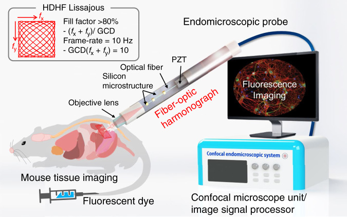
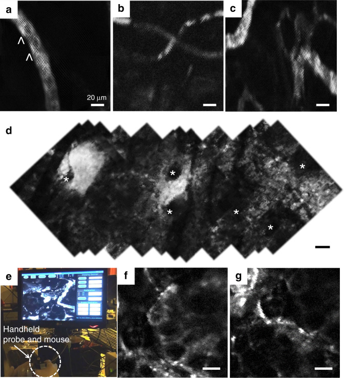
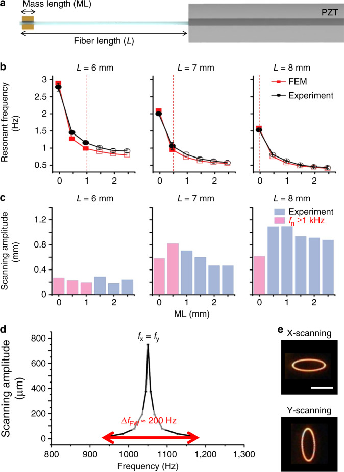
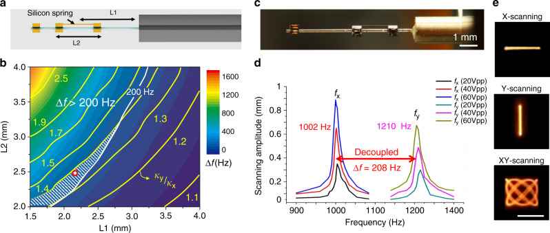
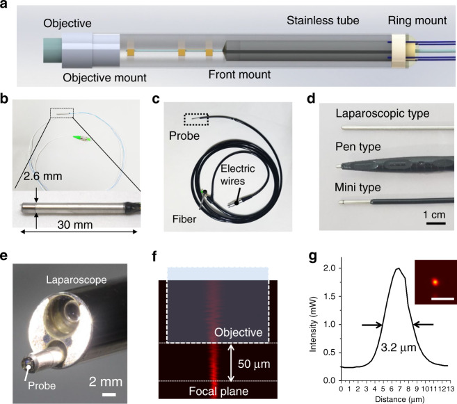
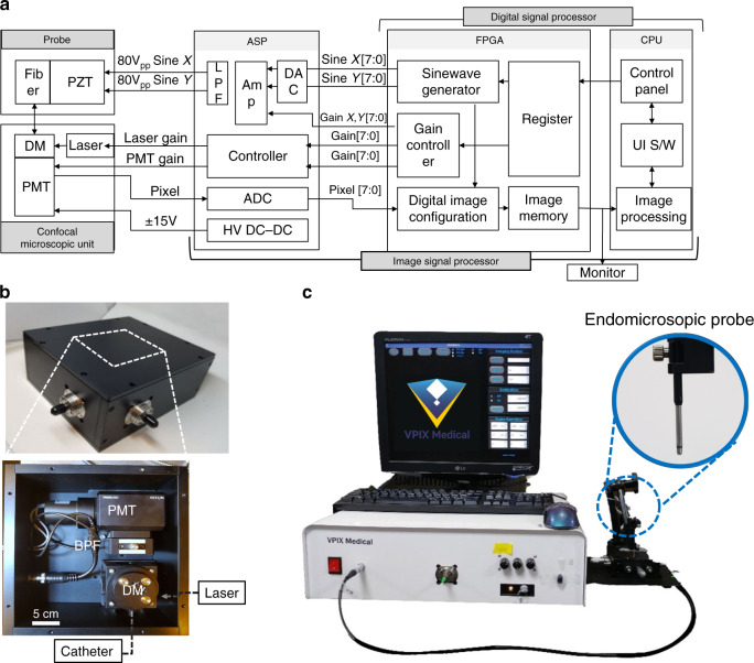
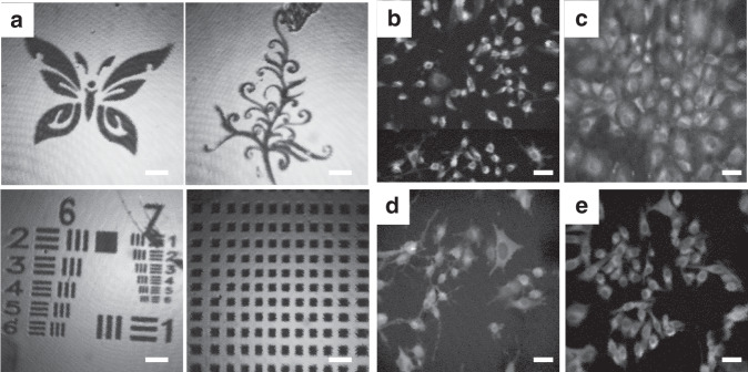
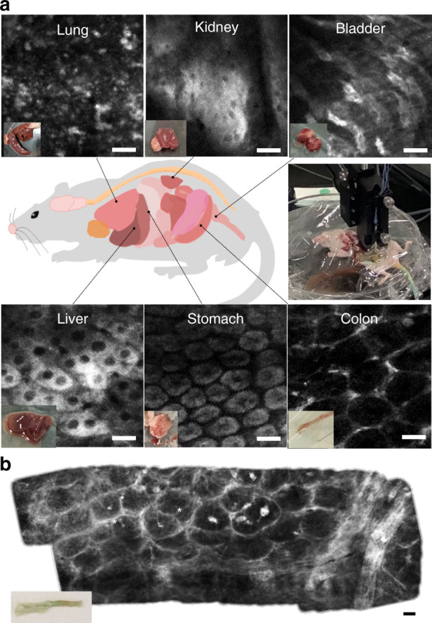
Confocal laser endomicroscopy provides high potential for noninvasive and in vivo optical biopsy at the cellular level. Here, we report a fully packaged handheld confocal endomicroscopic system for real-time, high-resolution, and in vivo cellular imaging using a Lissajous scanning fiber-optic harmonograph. The endomicroscopic system features an endomicroscopic probe with a fiber-optic harmonograph, a confocal microscope unit, and an image signal processor. The fiber-optic harmonograph contains a single mode fiber coupled with a quadrupole piezoelectric tube, which resonantly scans both axes at ~ 1 kHz to obtain a Lissajous pattern. The fiber-optic harmonograph was fully packaged into an endomicroscopic probe with an objective lens. The endomicroscopic probe was hygienically packaged for waterproofing and disinfection of medical instruments within a 2.6-mm outer diameter stainless tube capable of being inserted through the working channel of a clinical endoscope. The probe was further combined with the confocal microscope unit for indocyanine green imaging and the image signal processor for high frame rate and high density Lissajous scanning. The signal processing unit delivers driving signals for probe actuation and reconstructs confocal images using the auto phase matching process of Lissajous fiber scanners. The confocal endomicroscopic system was used to successfully obtain human in vitro fluorescent images and real-time ex vivo and in vivo fluorescent images of the living cell morphology and capillary perfusion inside a single mouse.
共聚焦激光内镜检查在细胞水平的无创和体内光学活检方面具有很高的潜力。在此,我们报告一种完全封装的手持式共聚焦内镜系统,该系统使用李萨如扫描光纤谐波记录仪进行实时、高分辨率的体内细胞成像。该内镜系统包括一个带有光纤谐波记录仪的内镜探头、一个共聚焦显微镜单元和一个图像信号处理器。光纤谐波记录仪包含一根与四极压电管耦合的单模光纤,该压电管以约1kHz的频率在两个轴上进行共振扫描以获得李萨如图形。光纤谐波记录仪被完全封装在一个带有物镜的内镜探头中。内镜探头经过卫生封装,以便在一个外径为2.6毫米的不锈钢管内对医疗器械进行防水和消毒,该不锈钢管能够通过临床内窥镜的工作通道插入。该探头进一步与用于吲哚菁绿成像的共聚焦显微镜单元以及用于高帧率和高密度李萨如扫描的图像信号处理器相结合。信号处理单元提供用于探头驱动的信号,并使用李萨如光纤扫描仪的自动相位匹配过程重建共聚焦图像。该共聚焦内镜系统成功地获得了人体体外荧光图像以及小鼠体内活细胞形态和毛细血管灌注的实时离体和体内荧光图像。