Department of Bio and Brain Engineering, KAIST, Daejeon, 34141, Republic of Korea.
KAIST Institute of Health science and technology, Daejeon, 34141, Republic of Korea.
Sci Rep. 2019 Mar 5;9(1):3560. doi: 10.1038/s41598-019-38762-w.
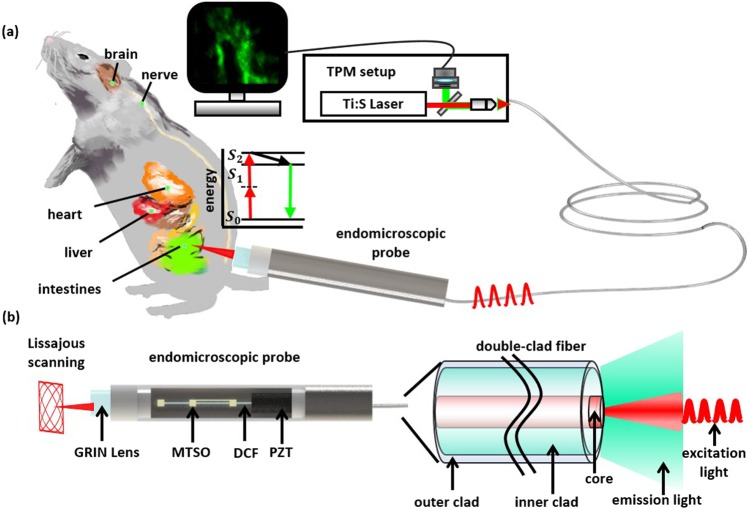
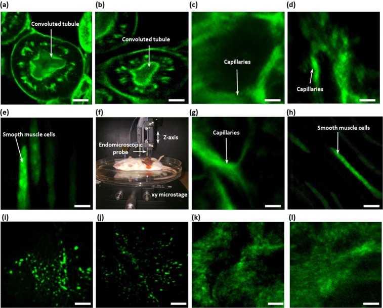
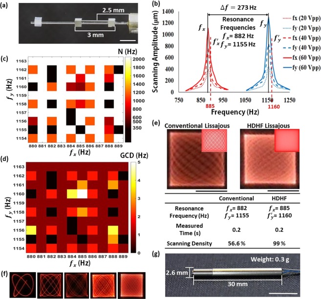
An endomicroscope opens new frontiers of non-invasive biopsy for in vivo imaging applications. Here we report two-photon laser scanning endomicroscope for in vivo cellular and tissue imaging using a Lissajous fiber scanner. The fiber scanner consists of a piezoelectric (PZT) tube, a single double-clad fiber (DCF) with high fluorescence collection, and a micro-tethered-silicon-oscillator (MTSO) for the separation of biaxial resonant scanning frequencies. The endomicroscopic imaging exhibits 5 frames/s with 99% in scanning density by using the selection rule of scanning frequencies. The endomicroscopic scanner was compactly packaged within a stainless tube of 2.6 mm in diameter with a high NA gradient-index (GRIN) lens, which can be easily inserted into the working channel of a conventional laparoscope. The lateral and axial resolutions of the endomicroscope are 0.70 µm and 7.6 μm, respectively. Two-photon fluorescence images of a stained kidney section and miscellaneous ex vivo and in vivo organs from wild type and green fluorescent protein transgenic (GFP-TG) mice were successfully obtained by using the endomicroscope. The endomicroscope also obtained label free images including autofluorescence and second-harmonic generation of an ear tissue of Thy1-GCaMP6 (GP5.17) mouse. The Lissajous scanning two-photon endomicroscope can provide a compact handheld platform for in vivo tissue imaging or optical biopsy applications.
内窥式显微镜为活体成像应用中的非侵入性活检开辟了新的领域。在这里,我们报告了一种使用双曲正弦光纤扫描仪的用于活体细胞和组织成像的双光子激光扫描内窥式显微镜。光纤扫描仪由一个压电(PZT)管、一个具有高荧光收集效率的单根双包层光纤(DCF)和一个用于分离双轴共振扫描频率的微栓硅振荡器(MTSO)组成。通过使用扫描频率的选择规则,内窥式显微镜以 5 帧/秒的速度实现了 99%的扫描密度。内窥式显微镜扫描仪被紧凑地封装在一个直径为 2.6 毫米的不锈钢管内,内置高数值孔径梯度折射率(GRIN)透镜,可轻松插入传统腹腔镜的工作通道。内窥式显微镜的横向和轴向分辨率分别为 0.70 μm 和 7.6 μm。通过使用内窥式显微镜,成功获得了染色肾切片以及野生型和绿色荧光蛋白转基因(GFP-TG)小鼠各种离体和体内器官的双光子荧光图像。内窥式显微镜还获得了包括 Thy1-GCaMP6(GP5.17)小鼠耳部组织的自发荧光和二次谐波产生的无标记图像。双曲正弦扫描双光子内窥式显微镜可以为活体组织成像或光学活检应用提供紧凑的手持式平台。