Chapin Ashley A, Rajasekaran Pradeep R, Quan David N, Hu Liangbing, Herberholz Jens, Bentley William E, Ghodssi Reza
Fischell Department of Bioengineering, College Park, MD 20742 USA.
Institute for Systems Research, College Park, MD 20740 USA.
Microsyst Nanoeng. 2020 Sep 7;6:90. doi: 10.1038/s41378-020-00184-4. eCollection 2020.
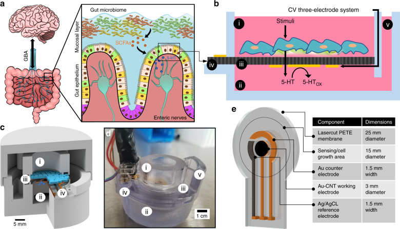
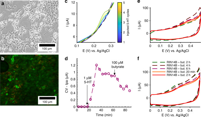
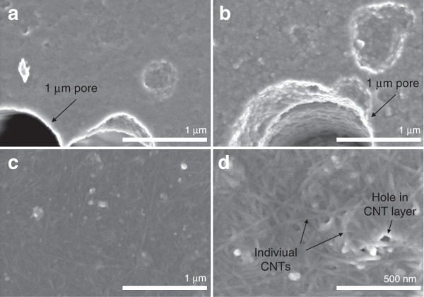
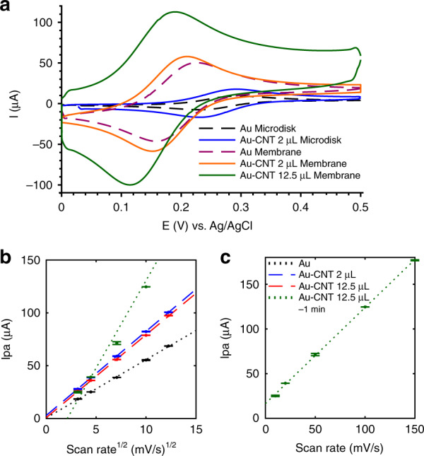
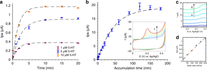
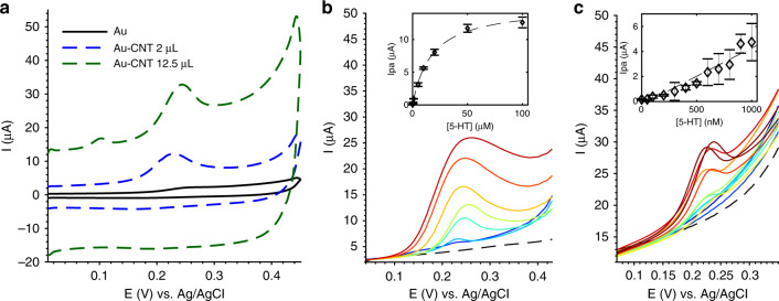
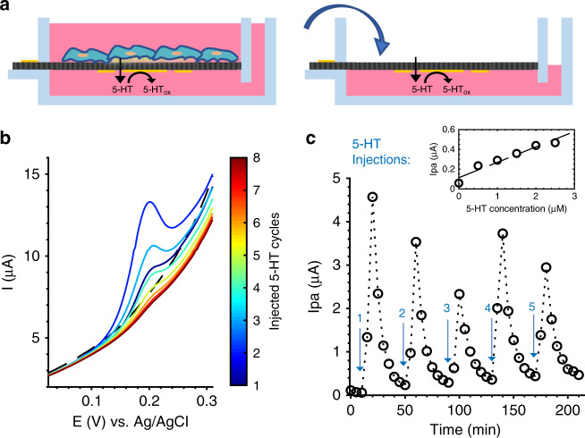
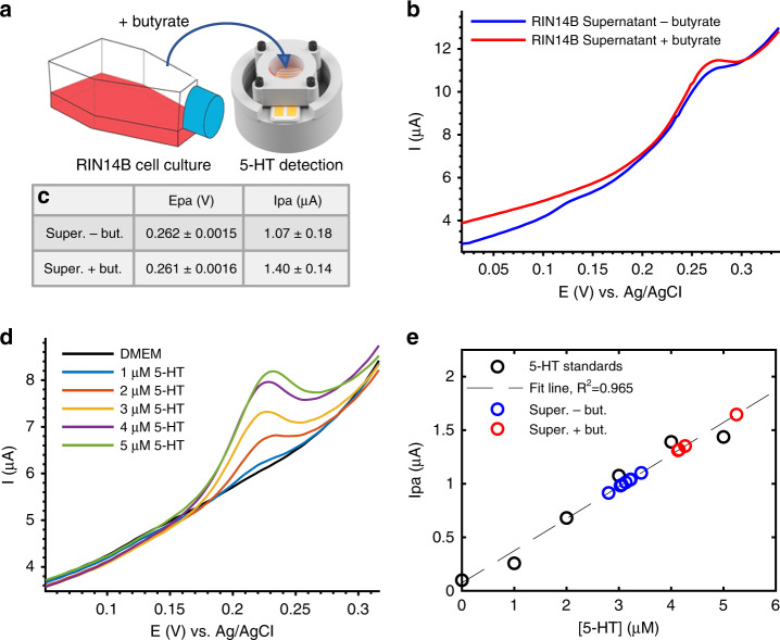
Gut-brain axis (GBA) communication relies on serotonin (5-HT) signaling between the gut epithelium and the peripheral nervous system, where 5-HT release patterns from the basolateral (i.e., bottom) side of the epithelium activate nerve afferents. There have been few quantitative studies of this gut-neuron signaling due to a lack of real-time measurement tools that can access the basolateral gut epithelium. In vitro platforms allow quantitative studies of cultured gut tissue, but they mainly employ offline and endpoint assays that cannot resolve dynamic molecular-release patterns. Here, we present the modification of a microporous cell culture membrane with carbon nanotube-coated gold (Au-CNT) electrodes capable of continuous, label-free, and direct detection of 5-HT at physiological concentrations. Electrochemical characterization of single-walled carbon nanotube (SWCNT)-coated Au electrodes shows increased electroactive surface area, 5-HT specificity, sensitivity, and saturation time, which are correlated with the CNT film drop-cast volume. Two microliters of CNT films, with a 10-min saturation time, 0.6 μA/μM 5-HT sensitivity, and reliable detection within a linear range of 500 nM-10 μM 5-HT, can be targeted for high-concentration, high-time-resolution 5-HT monitoring. CNT films (12.5 μL) with a 2-h saturation time, 4.5 μA/μM 5-HT sensitivity, and quantitative detection in the linear range of 100 nM-1 μM can target low concentrations with low time resolution. These electrodes achieved continuous detection of dynamic diffusion across the porous membrane, mimicking basolateral 5-HT release from cells, and detection of cell-released 5-HT from separately cultured RIN14B cell supernatant. Electrode-integrated cell culture systems such as this can improve in vitro molecular detection mechanisms and aid in quantitative GBA signaling studies.
肠-脑轴(GBA)通讯依赖于肠道上皮与外周神经系统之间的血清素(5-HT)信号传导,其中上皮基底外侧(即底部)的5-HT释放模式可激活神经传入纤维。由于缺乏能够接触肠道基底外侧上皮的实时测量工具,对这种肠-神经元信号传导的定量研究很少。体外平台可对培养的肠道组织进行定量研究,但主要采用离线和终点检测方法,无法解析动态分子释放模式。在此,我们展示了一种用碳纳米管包覆金(Au-CNT)电极对微孔细胞培养膜进行的改性,该电极能够连续、无标记且直接检测生理浓度的5-HT。单壁碳纳米管(SWCNT)包覆的金电极的电化学表征显示,其电活性表面积、5-HT特异性、灵敏度和饱和时间均有所增加,这与碳纳米管膜滴铸体积相关。两微升碳纳米管膜,饱和时间为10分钟,5-HT灵敏度为0.6 μA/μM,在500 nM - 10 μM的5-HT线性范围内可进行可靠检测,可用于高浓度、高时间分辨率的5-HT监测。饱和时间为2小时、5-HT灵敏度为4.5 μA/μM且在100 nM - 1 μM线性范围内进行定量检测的12.5 μL碳纳米管膜,可用于低时间分辨率的低浓度检测。这些电极实现了对跨多孔膜动态扩散的连续检测,模拟了细胞基底外侧5-HT的释放,并检测了单独培养的RIN14B细胞上清液中细胞释放的5-HT。这样的电极集成细胞培养系统可以改善体外分子检测机制,并有助于进行GBA信号传导的定量研究。