• 文献检索
  • 文档翻译
  • 深度研究
  • 学术资讯
  • Suppr Zotero 插件Zotero 插件
  • 邀请有礼
  • 套餐&价格
  • 历史记录
应用&插件
Suppr Zotero 插件Zotero 插件浏览器插件Mac 客户端Windows 客户端微信小程序
定价
高级版会员购买积分包购买API积分包
服务
文献检索文档翻译深度研究API 文档MCP 服务
关于我们
关于 Suppr公司介绍联系我们用户协议隐私条款
关注我们

Suppr 超能文献

核心技术专利:CN118964589B侵权必究
粤ICP备2023148730 号-1Suppr @ 2026

文献检索

告别复杂PubMed语法,用中文像聊天一样搜索,搜遍4000万医学文献。AI智能推荐,让科研检索更轻松。

立即免费搜索

文件翻译

保留排版,准确专业,支持PDF/Word/PPT等文件格式,支持 12+语言互译。

免费翻译文档

深度研究

AI帮你快速写综述,25分钟生成高质量综述,智能提取关键信息,辅助科研写作。

立即免费体验

使用超高通量全息细胞术对储存的红细胞进行单细胞分析。

Single Cell Analysis of Stored Red Blood Cells Using Ultra-High Throughput Holographic Cytometry.

机构信息

Department of Biomedical Engineering, Duke University, Durham, NC 27708, USA.

Department of Molecular Genetics and Microbiology, Duke University, Durham, NC 27708, USA.

出版信息

Cells. 2021 Sep 17;10(9):2455. doi: 10.3390/cells10092455.

DOI:10.3390/cells10092455
PMID:34572104
原文链接:https://pmc.ncbi.nlm.nih.gov/articles/PMC8465484/
Abstract

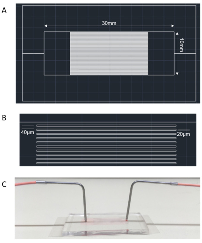
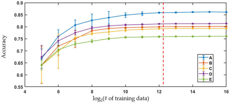
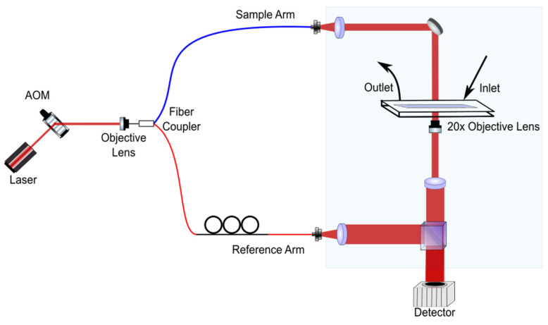
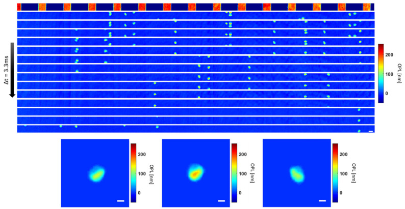
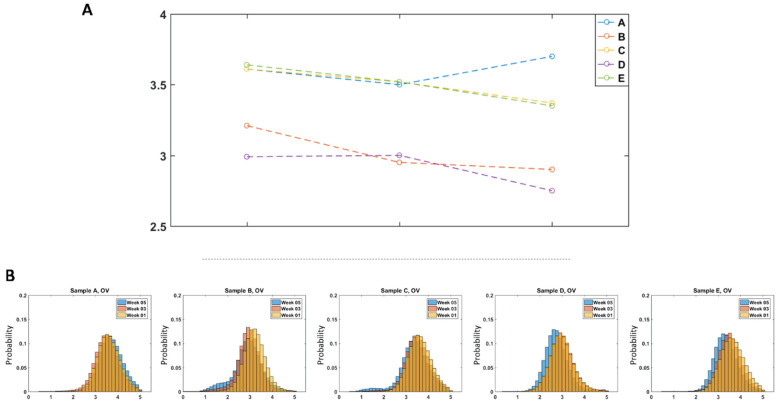
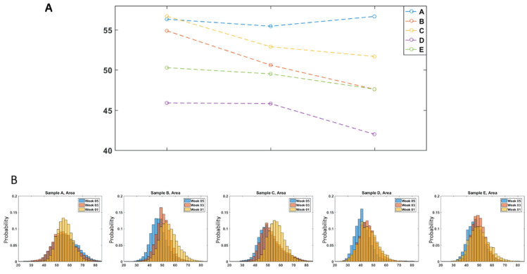
Holographic cytometry is introduced as an ultra-high throughput implementation of quantitative phase imaging of single cells flowing through parallel microfluidic channels. Here, the approach was applied for characterizing the morphology of individual red blood cells during storage under regular blood bank conditions. Samples from five blood donors were examined, over 100,000 cells examined for each, at three time points. The approach allows high-throughput phase imaging of a large number of cells, greatly extending our ability to study cellular phenotypes using individual cell images. Holographic cytology images can provide measurements of multiple physical traits of the cells, including optical volume and area, which are observed to consistently change over the storage time. In addition, the large volume of cell imaging data can serve as training data for machine-learning algorithms. For the study here, logistic regression was used to classify the cells according to the storage time points. The analysis showed that at least 5000 cells are needed to ensure accuracy of the classifiers. Overall, results showed the potential of holographic cytometry as a diagnostic tool.

摘要

体全息细胞术作为一种超高速高通量的单细胞定量相位成像方法被引入。在这里,该方法被应用于在常规血库条件下储存过程中单个红细胞形态的特征化研究。对五名献血者的样本进行了检查,每个样本检查了超过 100,000 个细胞,在三个时间点进行了检查。该方法允许对大量细胞进行高通量相位成像,极大地扩展了我们使用单个细胞图像研究细胞表型的能力。体全息细胞术图像可以提供细胞的多个物理特征的测量值,包括光学体积和面积,这些特征在储存时间内观察到持续变化。此外,大量的细胞成像数据可以作为机器学习算法的训练数据。在本研究中,逻辑回归被用于根据储存时间点对细胞进行分类。分析表明,至少需要 5000 个细胞才能确保分类器的准确性。总体而言,结果表明体全息细胞术作为一种诊断工具具有潜力。

https://cdn.ncbi.nlm.nih.gov/pmc/blobs/74f4/8465484/b1ff03f5a537/cells-10-02455-g007.jpg
https://cdn.ncbi.nlm.nih.gov/pmc/blobs/74f4/8465484/61f2a5369e6a/cells-10-02455-g001.jpg
https://cdn.ncbi.nlm.nih.gov/pmc/blobs/74f4/8465484/0df0ac70092c/cells-10-02455-g002.jpg
https://cdn.ncbi.nlm.nih.gov/pmc/blobs/74f4/8465484/8d71fc7584e4/cells-10-02455-g003.jpg
https://cdn.ncbi.nlm.nih.gov/pmc/blobs/74f4/8465484/1cebbdfd471d/cells-10-02455-g004.jpg
https://cdn.ncbi.nlm.nih.gov/pmc/blobs/74f4/8465484/da067d2a6c93/cells-10-02455-g005.jpg
https://cdn.ncbi.nlm.nih.gov/pmc/blobs/74f4/8465484/7b507e4be0fa/cells-10-02455-g006.jpg
https://cdn.ncbi.nlm.nih.gov/pmc/blobs/74f4/8465484/b1ff03f5a537/cells-10-02455-g007.jpg

相似文献

1
Single Cell Analysis of Stored Red Blood Cells Using Ultra-High Throughput Holographic Cytometry.使用超高通量全息细胞术对储存的红细胞进行单细胞分析。
Cells. 2021 Sep 17;10(9):2455. doi: 10.3390/cells10092455.
2
Accurate label-free 3-part leukocyte recognition with single cell lens-free imaging flow cytometry.使用无镜头的流式细胞术对单细胞进行无标记的 3 部分白细胞识别。
Comput Biol Med. 2018 May 1;96:147-156. doi: 10.1016/j.compbiomed.2018.03.008. Epub 2018 Mar 14.
3
Three-dimensional quantitative phase imaging of blood coagulation structures by optical projection tomography in flow cytometry using digital holographic microscopy.采用数字全息显微镜的流式细胞术光学投影断层成像术对血液凝固结构的三维定量相位成像。
J Biomed Opt. 2018 Oct;24(3):1-6. doi: 10.1117/1.JBO.24.3.031012.
4
Screening for urothelial carcinoma cells in urine based on digital holographic flow cytometry through machine learning and deep learning methods.基于机器学习和深度学习方法的数字全息流式细胞术在尿液中检测尿路上皮癌细胞。
Lab Chip. 2024 May 14;24(10):2736-2746. doi: 10.1039/d3lc00854a.
5
Label-free, high-throughput holographic screening and enumeration of tumor cells in blood.无标记、高通量全息筛选和血液中肿瘤细胞的计数。
Lab Chip. 2017 Aug 22;17(17):2920-2932. doi: 10.1039/c7lc00149e.
6
Automated quantitative analysis of 3D morphology and mean corpuscular hemoglobin in human red blood cells stored in different periods.不同储存时期人红细胞三维形态及平均红细胞血红蛋白的自动化定量分析
Opt Express. 2013 Dec 16;21(25):30947-57. doi: 10.1364/OE.21.030947.
7
A practical criterion for focusing of unstained cell samples using a digital holographic microscope.使用数字全息显微镜聚焦未染色细胞样品的实用标准。
J Microsc. 2020 Aug;279(2):114-122. doi: 10.1111/jmi.12924. Epub 2020 Jun 5.
8
Predicting single-cell gene expression profiles of imaging flow cytometry data with machine learning.利用机器学习预测成像流式细胞术数据的单细胞基因表达谱。
Nucleic Acids Res. 2020 Nov 18;48(20):11335-11346. doi: 10.1093/nar/gkaa926.
9
Design and development of volume phase holographic grating based digital holographic interferometer for label-free quantitative cell imaging.基于体相位全息光栅的数字全息干涉仪的设计与开发,用于无标记定量细胞成像。
Appl Opt. 2020 Apr 20;59(12):3773-3783. doi: 10.1364/AO.387620.
10
High accuracy label-free classification of single-cell kinetic states from holographic cytometry of human melanoma cells.高准确度的无标记单细胞动力学状态分类,基于人类黑色素瘤细胞的全息细胞术。
Sci Rep. 2017 Sep 20;7(1):11943. doi: 10.1038/s41598-017-12165-1.

引用本文的文献

1
New and emerging technologies for pretransfusion blood quality assessment: A state-of-the-art review.输血前血液质量评估的新兴技术:最新综述。
Transfusion. 2024 Nov;64(11):2196-2208. doi: 10.1111/trf.18019. Epub 2024 Sep 26.
2
Perspective on quantitative phase imaging to improve precision cancer medicine.定量相位成像在提高精准癌症医学中的应用展望。
J Biomed Opt. 2024 Jun;29(Suppl 2):S22705. doi: 10.1117/1.JBO.29.S2.S22705. Epub 2024 Mar 26.
3
Biophysical Profiling of Sickle Cell Disease Using Holographic Cytometry and Deep Learning.

本文引用的文献

1
Quantitative phase imaging of erythrocytes under microfluidic constriction in a high refractive index medium reveals water content changes.在高折射率介质中微流体收缩条件下对红细胞进行定量相成像揭示了水分含量变化。
Microsyst Nanoeng. 2019 Dec 2;5:63. doi: 10.1038/s41378-019-0113-y. eCollection 2019.
2
Invited Article: Digital refocusing in quantitative phase imaging for flowing red blood cells.特邀文章:流动红细胞定量相成像中的数字重聚焦
APL Photonics. 2018 Nov;3(11). doi: 10.1063/1.5043536. Epub 2018 Oct 2.
3
Red blood cell storage lesion: causes and potential clinical consequences.
使用全息细胞术和深度学习对镰状细胞病进行生物物理分析。
Int J Mol Sci. 2023 Jul 25;24(15):11885. doi: 10.3390/ijms241511885.
4
Multiscale optical phase fluctuations link disorder strength and fractal dimension of cell structure.多尺度光学相位涨落与细胞结构的无序强度和分形维数有关。
Biophys J. 2023 Apr 4;122(7):1390-1399. doi: 10.1016/j.bpj.2023.03.005. Epub 2023 Mar 5.
5
Microfluidic Systems for Blood and Blood Cell Characterization.微流控系统在血液和血细胞分析中的应用。
Biosensors (Basel). 2022 Dec 22;13(1):13. doi: 10.3390/bios13010013.
6
Single-Cell Analysis 2.0.单细胞分析 2.0.
Cells. 2022 Dec 30;12(1):154. doi: 10.3390/cells12010154.
7
Cutting-Edge Methods for Better Understanding Cells.用于更好地理解细胞的尖端方法。
Cells. 2022 Nov 3;11(21):3479. doi: 10.3390/cells11213479.
8
Predicting willingness to donate blood based on machine learning: two blood donor recruitments during COVID-19 outbreaks.基于机器学习预测献血意愿:COVID-19 疫情期间的两次献血者招募。
Sci Rep. 2022 Nov 10;12(1):19165. doi: 10.1038/s41598-022-21215-2.
9
Erysense, a Lab-on-a-Chip-Based Point-of-Care Device to Evaluate Red Blood Cell Flow Properties With Multiple Clinical Applications.Erysense,一种基于芯片实验室的即时检测设备,用于评估红细胞流动特性并具有多种临床应用。
Front Physiol. 2022 Apr 27;13:884690. doi: 10.3389/fphys.2022.884690. eCollection 2022.
10
Single Cell RNA-Seq Analysis of Human Red Cells.人类红细胞的单细胞RNA测序分析
Front Physiol. 2022 Apr 20;13:828700. doi: 10.3389/fphys.2022.828700. eCollection 2022.
红细胞储存损伤:原因及潜在临床后果。
Blood Transfus. 2019 Jan;17(1):27-52. doi: 10.2450/2019.0217-18.
4
Angiogenin-mediated tRNA cleavage as a novel feature of stored red blood cells.血管生成素介导的tRNA切割作为储存红细胞的一个新特征。
Br J Haematol. 2019 May;185(4):760-764. doi: 10.1111/bjh.15605. Epub 2018 Oct 18.
5
Complete Blood Count Reference Intervals and Patterns of Changes Across Pediatric, Adult, and Geriatric Ages in Korea.韩国儿科、成人和老年人群的全血细胞计数参考区间和变化模式。
Ann Lab Med. 2018 Nov;38(6):503-511. doi: 10.3343/alm.2018.38.6.503.
6
Red blood cells ageing markers: a multi-parametric analysis.红细胞衰老标志物:多参数分析
Blood Transfus. 2017 May;15(3):239-248. doi: 10.2450/2017.0318-16.
7
Optical Phase Measurements of Disorder Strength Link Microstructure to Cell Stiffness.无序强度的光学相位测量将微观结构与细胞刚度联系起来。
Biophys J. 2017 Feb 28;112(4):692-702. doi: 10.1016/j.bpj.2016.12.016.
8
Spherocytic shift of red blood cells during storage provides a quantitative whole cell-based marker of the storage lesion.储存期间红细胞的球形转变提供了一种基于全细胞的储存损伤定量标志物。
Transfusion. 2017 Apr;57(4):1007-1018. doi: 10.1111/trf.14015. Epub 2017 Feb 1.
9
Review: imaging technologies for flow cytometry.综述:流式细胞术的成像技术。
Lab Chip. 2016 Nov 29;16(24):4639-4647. doi: 10.1039/c6lc01063f.
10
Automated Detection of P. falciparum Using Machine Learning Algorithms with Quantitative Phase Images of Unstained Cells.使用机器学习算法和未染色细胞的定量相位图像自动检测恶性疟原虫。
PLoS One. 2016 Sep 16;11(9):e0163045. doi: 10.1371/journal.pone.0163045. eCollection 2016.