Jeon Ji-Hyeon, Lee Jaehyeok, Park Jin-Hyang, Lee Chul-Haeng, Choi Min-Koo, Song Im-Sook
BK21 FOUR Community-Based Intelligent Novel Drug Discovery Education Unit, Vessel-Organ Interaction Research Center (VOICE), Research Institute of Pharmaceutical Sciences, College of Pharmacy, Kyungpook National University, Daegu 41566, Korea.
College of Pharmacy, Dankook University, Cheon-an 31116, Korea.
Pharmaceutics. 2021 Sep 17;13(9):1496. doi: 10.3390/pharmaceutics13091496.
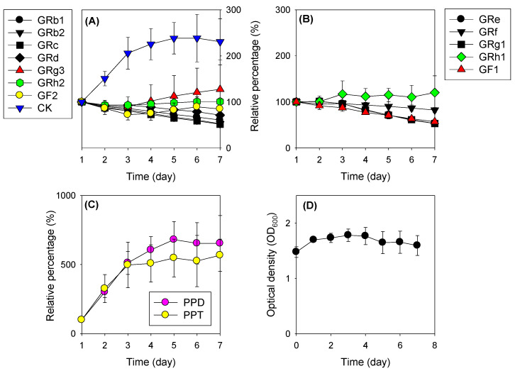
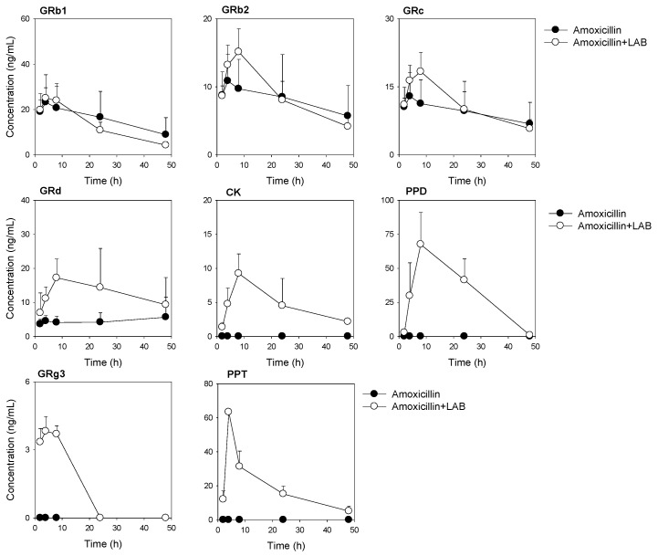
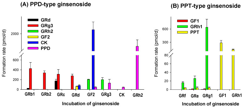
This study aims to investigate the effect of lactic acid bacteria (LAB) on in vitro and in vivo metabolism and the pharmacokinetics of ginsenosides in mice. When the in vitro fermentation test of RGE with LAB was carried out, protopanaxadiol (PPD) and protopanaxadiol (PPD), which are final metabolites of ginsenosides but not contained in RGE, were greatly increased. Compound K (CK), ginsenoside Rh1 (GRh1), and GRg3 also increased by about 30%. Other ginsenosides with a sugar number of more than 2 showed a gradual decrease by fermentation with LAB for 7 days, suggesting the involvement of LAB in the deglycosylation of ginsenosides. Incubation of single ginsenoside with LAB produced GRg3, CK, and PPD with the highest formation rate and GRd, GRh2, and GF with the lower rate among PPD-type ginsenosides. Among PPT-type ginsenosides, GRh1 and PPT had the highest formation rate. The amoxicillin pretreatment (20 mg/kg/day, twice a day for 3 days) resulted in a significant decrease in the fecal recovery of CK, PPD, and PPT through the blockade of deglycosylation of ginsenosides after single oral administrations of RGE (2 g/kg) in mice. The plasma concentrations of CK, PPD, and PPT were not detectable without change in GRb1, GRb2, and GRc in this group. LAB supplementation (1 billion CFU/2 g/kg/day for 1 week) after the amoxicillin treatment in mice restored the ginsenoside metabolism and the plasma concentrations of ginsenosides to the control level. In conclusion, the alterations in the gut microbiota environment could change the ginsenoside metabolism and plasma concentrations of ginsenosides. Therefore, the supplementation of LAB with oral administrations of RGE would help increase plasma concentrations of deglycosylated ginsenosides such as CK, PPD, and PPT.
本研究旨在探讨乳酸菌(LAB)对小鼠体内外代谢及人参皂苷药代动力学的影响。在用LAB对红参提取物(RGE)进行体外发酵试验时,人参皂苷的最终代谢产物原人参二醇(PPD)和原人参三醇(PPT)(RGE中不含)大幅增加。化合物K(CK)、人参皂苷Rh1(GRh1)和GRg3也增加了约30%。其他糖基数超过2的人参皂苷经LAB发酵7天后逐渐减少,表明LAB参与了人参皂苷的去糖基化过程。单一人参皂苷与LAB孵育产生GRg3、CK和PPD的生成率最高,而在PPD型人参皂苷中,GRd、GRh2和GF的生成率较低。在PPT型人参皂苷中,GRh1和PPT的生成率最高。阿莫西林预处理(20毫克/千克/天,每天两次,共3天)导致小鼠单次口服RGE(2克/千克)后,通过阻断人参皂苷的去糖基化,CK、PPD和PPT的粪便回收率显著降低。该组中未检测到CK、PPD和PPT的血浆浓度,而GRb1、GRb2和GRc无变化。小鼠经阿莫西林治疗后补充LAB(10亿CFU/2克/千克/天,共1周)可使人参皂苷代谢和人参皂苷血浆浓度恢复至对照水平。总之,肠道微生物群环境的改变可改变人参皂苷代谢和人参皂苷血浆浓度。因此,口服RGE时补充LAB有助于提高CK、PPD和PPT等去糖基化人参皂苷的血浆浓度。