Section of Virology, Faculty of Veterinary Medicine, Norwegian University of Life Sciences, Ås, Norway.
Department of Fish Health, Norwegian Veterinary Institute, Ås, Norway.
Front Immunol. 2021 Sep 16;12:729017. doi: 10.3389/fimmu.2021.729017. eCollection 2021.
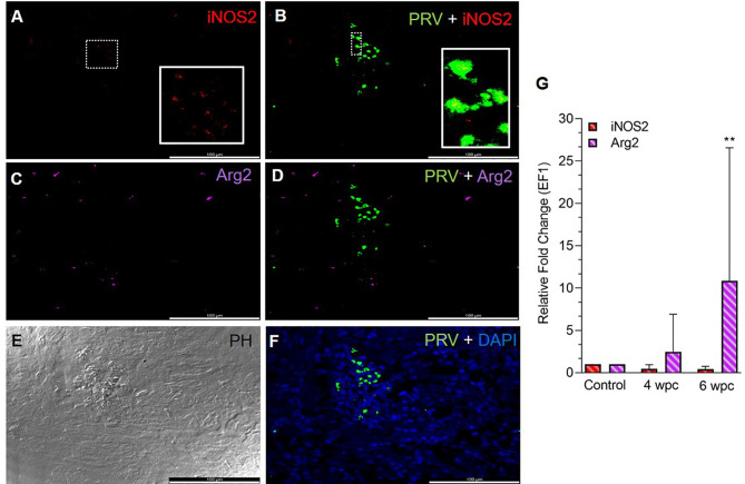
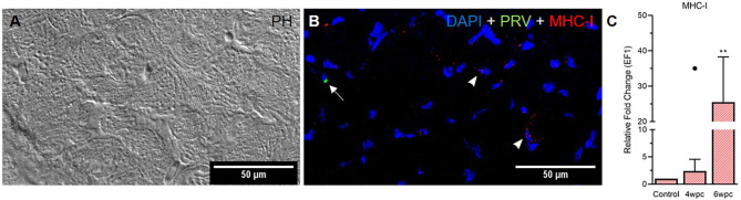
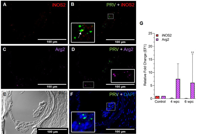
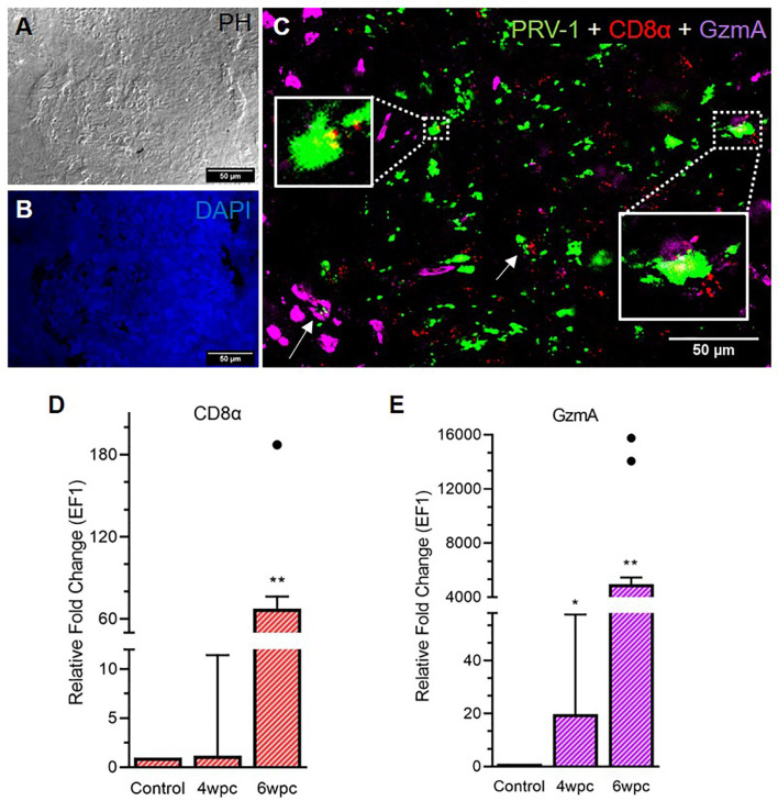
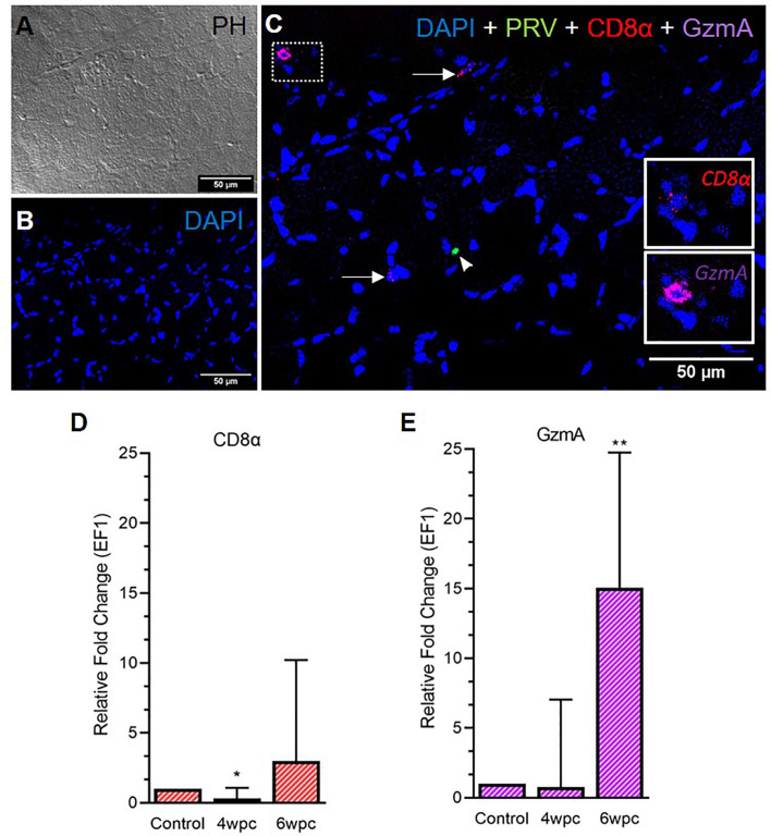
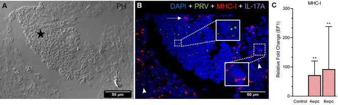
Piscine orthoreovirus (PRV-1) infection causes heart and skeletal muscle inflammation (HSMI) in farmed Atlantic salmon . The virus is also associated with focal melanized changes in white skeletal muscle where PRV-1 infection of macrophages appears to be important. In this study, we studied the macrophage polarization into M1 (pro-inflammatory) and M2 (anti-inflammatory) phenotypes during experimentally induced HSMI. The immune response in heart with HSMI lesions was characterized by CD8 and MHC-I expressing cells and not by polarized macrophages. Fluorescent hybridization (FISH) assays revealed localization of PRV-1 in a few M1 macrophages in both heart and skeletal muscle. M2 type macrophages were widely scattered in the heart and were more abundant in heart compared to the skeletal muscle. However, the M2 macrophages did not co-stain for PRV-1. There was a strong cellular immune response to the infection in the heart compared to that of the skeletal muscle, seen as increased MHC-I expression, partly in cells also containing PRV-1 RNA, and a high number of cytotoxic CD8 granzyme producing cells that targeted PRV-1. In skeletal muscle, MHC-I expressing cells and CD8 cells were dispersed between myocytes, but these cells did not stain for PRV-1. Gene expression analysis by RT-qPCR complied with the FISH results and confirmed a drop in level of PRV-1 following the cell mediated immune response. Overall, the results indicated that M1 macrophages do not contribute to the initial development of HSMI. However, large numbers of M2 macrophages reside in the heart and may contribute to the subsequent fast recovery following clearance of PRV-1 infection.
鱼类正呼肠孤病毒(PRV-1)感染会引起养殖大西洋鲑的心肌和骨骼肌炎症(HSMI)。该病毒还与白色骨骼肌中的局灶性黑化变化有关,PRV-1 对巨噬细胞的感染似乎很重要。在这项研究中,我们研究了在实验诱导的 HSMI 期间巨噬细胞向 M1(促炎)和 M2(抗炎)表型的极化。具有 HSMI 病变的心脏中的免疫反应特征是 CD8 和 MHC-I 表达细胞,而不是极化的巨噬细胞。荧光杂交(FISH)检测显示 PRV-1 定位于心脏和骨骼肌中的少数 M1 巨噬细胞中。M2 型巨噬细胞广泛分布在心脏中,在心脏中的丰度高于骨骼肌。然而,M2 巨噬细胞没有与 PRV-1 共染色。与骨骼肌相比,心脏对感染的细胞免疫反应更强,表现为 MHC-I 表达增加,部分细胞中也含有 PRV-1 RNA,以及大量靶向 PRV-1 的细胞毒性 CD8 颗粒酶产生细胞。在骨骼肌中,MHC-I 表达细胞和 CD8 细胞散布在肌细胞之间,但这些细胞没有与 PRV-1 染色。RT-qPCR 基因表达分析与 FISH 结果一致,并证实细胞介导的免疫反应后 PRV-1 水平下降。总体而言,结果表明 M1 巨噬细胞不会导致 HSMI 的初始发展。然而,大量的 M2 巨噬细胞存在于心脏中,可能有助于 PRV-1 感染清除后的快速恢复。