• 文献检索
  • 文档翻译
  • 深度研究
  • 学术资讯
  • Suppr Zotero 插件Zotero 插件
  • 邀请有礼
  • 套餐&价格
  • 历史记录
应用&插件
Suppr Zotero 插件Zotero 插件浏览器插件Mac 客户端Windows 客户端微信小程序
定价
高级版会员购买积分包购买API积分包
服务
文献检索文档翻译深度研究API 文档MCP 服务
关于我们
关于 Suppr公司介绍联系我们用户协议隐私条款
关注我们

Suppr 超能文献

核心技术专利:CN118964589B侵权必究
粤ICP备2023148730 号-1Suppr @ 2026

文献检索

告别复杂PubMed语法,用中文像聊天一样搜索,搜遍4000万医学文献。AI智能推荐,让科研检索更轻松。

立即免费搜索

文件翻译

保留排版,准确专业,支持PDF/Word/PPT等文件格式,支持 12+语言互译。

免费翻译文档

深度研究

AI帮你快速写综述,25分钟生成高质量综述,智能提取关键信息,辅助科研写作。

立即免费体验

通过 OCT 血管造影定量分析,探讨毛细血管充血与慢性分支视网膜静脉阻塞黄斑水肿复发的关系。

Association between capillary congestion and macular edema recurrence in chronic branch retinal vein occlusion through quantitative analysis of OCT angiography.

机构信息

Department of Ophthalmology, Pusan National University Yangsan Hospital, Geumo-ro 20, Mulgeum-eup, Yangsan-si, Gyeongsangnam-do, 50612, South Korea.

Research Institute for Convergence of Biomedical Science and Technology, Pusan National University Yangsan Hospital, Geumo-ro 20, Mulgeum-eup, Yangsan-si, Gyeongsangnam-do, 50612, South Korea.

出版信息

Sci Rep. 2021 Oct 6;11(1):19886. doi: 10.1038/s41598-021-99429-z.

DOI:10.1038/s41598-021-99429-z
PMID:34615979
原文链接:https://pmc.ncbi.nlm.nih.gov/articles/PMC8494742/
Abstract

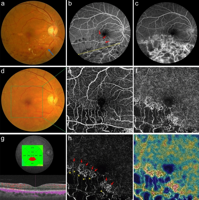
This study aims to quantitatively investigate the optical coherence tomographic angiography (OCTA) findings of capillary congestion and its association with macular edema (ME) recurrence in chronic branch retinal vein occlusion (BRVO). We retrospectively reviewed the medical records of 115 consecutive patients with major ischemic BRVO who reached stable macula (without ME for two consecutive visits) at baseline (the first visit within the stable period). All patients were classified into a recurrence or non-recurrence groups depending on ME recurrence. Capillary congestion of deep capillary plexuses (DCP-C) and other abnormal capillary lesions were segmented, and their areas, vascular densities, and mean retinal thicknesses (MRT) were calculated. The main outcomes were differences between the two groups and risk factors for recurrence among baseline and OCTA parameters. A total of 76 eyes were included, of which 22 (28.9%) recurred. DCP-C existed in all eyes at baseline. MRT of DCP-C (p = 0.006) was greater in the recurrence group. Greater MRT of DCP-C (OR: 1.044; p = 0.002) and more frequent intravitreal injections (OR: 1.803; p < 0.001) were associated with a higher risk of relapsing ME. DCP-C may contribute to the anatomical stability of chronic BRVO and simultaneously be the source of ME.

摘要

本研究旨在定量研究毛细血管充血的光相干断层扫描血管造影 (OCTA) 表现及其与慢性分支视网膜静脉阻塞 (BRVO) 黄斑水肿 (ME) 复发的关系。我们回顾性分析了 115 例主要缺血性 BRVO 连续患者的病历,这些患者在基线时(稳定期内的首次就诊)达到稳定的黄斑(连续两次就诊均无 ME)。所有患者根据 ME 复发情况分为复发组或非复发组。对深层毛细血管丛 (DCP-C) 的毛细血管充血和其他异常毛细血管病变进行分割,并计算其面积、血管密度和平均视网膜厚度 (MRT)。主要结果是两组之间的差异以及基线和 OCTA 参数中复发的危险因素。共纳入 76 只眼,其中 22 只(28.9%)复发。基线时所有眼均存在 DCP-C。复发组 DCP-C 的 MRT 较大(p=0.006)。DCP-C 的更大 MRT(OR:1.044;p=0.002)和更频繁的玻璃体内注射(OR:1.803;p<0.001)与 ME 复发的风险增加相关。DCP-C 可能有助于慢性 BRVO 的解剖学稳定,同时也是 ME 的来源。

https://cdn.ncbi.nlm.nih.gov/pmc/blobs/05b1/8494742/49d4a6e1c8a5/41598_2021_99429_Fig4_HTML.jpg
https://cdn.ncbi.nlm.nih.gov/pmc/blobs/05b1/8494742/c3e3bfa1a5fc/41598_2021_99429_Fig1_HTML.jpg
https://cdn.ncbi.nlm.nih.gov/pmc/blobs/05b1/8494742/6f2f2d84484b/41598_2021_99429_Fig2_HTML.jpg
https://cdn.ncbi.nlm.nih.gov/pmc/blobs/05b1/8494742/a4055e29c3cc/41598_2021_99429_Fig3_HTML.jpg
https://cdn.ncbi.nlm.nih.gov/pmc/blobs/05b1/8494742/49d4a6e1c8a5/41598_2021_99429_Fig4_HTML.jpg

相似文献

1
Association between capillary congestion and macular edema recurrence in chronic branch retinal vein occlusion through quantitative analysis of OCT angiography.通过 OCT 血管造影定量分析,探讨毛细血管充血与慢性分支视网膜静脉阻塞黄斑水肿复发的关系。
Sci Rep. 2021 Oct 6;11(1):19886. doi: 10.1038/s41598-021-99429-z.
2
OCT angiography features associated with macular edema recurrence after intravitreal bevacizumab treatment in branch retinal vein occlusion.OCT 血管造影特征与分支视网膜静脉阻塞玻璃体内注射贝伐单抗治疗后黄斑水肿复发的关系。
Sci Rep. 2019 Oct 2;9(1):14153. doi: 10.1038/s41598-019-50637-8.
3
Correlation between macular edema recurrence and macular capillary network destruction in branch retinal vein occlusion.视网膜分支静脉阻塞中黄斑水肿复发与黄斑毛细血管网络破坏之间的相关性
BMC Ophthalmol. 2020 Aug 24;20(1):341. doi: 10.1186/s12886-020-01611-w.
4
Grading of macular perfusion in retinal vein occlusion using en-face swept-source optical coherence tomography angiography: a retrospective observational case series.使用表面扫频源光学相干断层扫描血管造影术对视网膜静脉阻塞中的黄斑灌注进行分级:一项回顾性观察病例系列研究。
BMC Ophthalmol. 2019 Jun 10;19(1):127. doi: 10.1186/s12886-019-1134-x.
5
Gap in Capillary Perfusion on Optical Coherence Tomography Angiography Associated With Persistent Macular Edema in Branch Retinal Vein Occlusion.光学相干断层扫描血管造影显示的视网膜分支静脉阻塞伴持续性黄斑水肿患者的毛细血管灌注缺损
Invest Ophthalmol Vis Sci. 2017 Apr 1;58(4):2038-2043. doi: 10.1167/iovs.17-21447.
6
Optical coherence tomography angiography findings in patients with branch retinal vein occlusion treated with Anti-VEGF.接受抗血管内皮生长因子治疗的视网膜分支静脉阻塞患者的光学相干断层扫描血管造影结果
Arq Bras Oftalmol. 2020 Mar-Apr;83(2):120-126. doi: 10.5935/0004-2749.20200017.
7
Quantification of retinal microvasculature and neurodegeneration changes in branch retinal vein occlusion after resolution of cystoid macular edema on optical coherence tomography angiography.在光学相干断层扫描血管造影检查发现并解决了囊样黄斑水肿后,对视网膜分支静脉阻塞的视网膜微血管和神经退行性变变化进行定量分析。
Indian J Ophthalmol. 2019 Nov;67(11):1864-1869. doi: 10.4103/ijo.IJO_1554_18.
8
Optical Coherence Tomography Angiography Analysis of the Foveal Avascular Zone and Macular Vessel Density After Anti-VEGF Therapy in Eyes With Diabetic Macular Edema and Retinal Vein Occlusion.糖尿病性黄斑水肿和视网膜静脉阻塞患者抗VEGF治疗后黄斑无血管区和黄斑血管密度的光学相干断层扫描血管造影分析
Invest Ophthalmol Vis Sci. 2017 Jan 1;58(1):30-34. doi: 10.1167/iovs.16-20579.
9
[Anatomo-functional study in branch retinal vein occlusion using Swept Source Optical Coherence Tomography Angiography].[使用扫频源光学相干断层扫描血管造影术对视网膜分支静脉阻塞进行的解剖功能研究]
J Fr Ophtalmol. 2019 Mar;42(3):255-261. doi: 10.1016/j.jfo.2018.09.010. Epub 2019 Mar 8.
10
NOVEL OPTICAL COHERENCE TOMOGRAPHY ANGIOGRAPHY BIOMARKER IN BRANCH RETINAL VEIN OCCLUSION MACULAR EDEMA.新型光学相干断层扫描血管造影生物标志物在分支型视网膜静脉阻塞黄斑水肿中的应用。
Retina. 2019 Oct;39(10):1906-1916. doi: 10.1097/IAE.0000000000002264.

引用本文的文献

1
Soul: An OCTA dataset based on Human Machine Collaborative Annotation Framework.灵魂:基于人机协作标注框架的 OCTA 数据集。
Sci Data. 2024 Aug 2;11(1):838. doi: 10.1038/s41597-024-03665-7.
2
Role of optical coherence tomography angiography in the evaluation of peripheral ischemia in retinal vein occlusion.光学相干断层扫描血管造影在视网膜静脉阻塞外周缺血评估中的作用。
Saudi J Ophthalmol. 2024 Feb 23;38(2):138-143. doi: 10.4103/sjopt.sjopt_182_23. eCollection 2024 Apr-Jun.
3
Alteration of perivascular reflectivity on optical coherence tomography of branched retinal vein obstruction.

本文引用的文献

1
Associations Between Capillary Diameter, Capillary Density, and Microaneurysms in Diabetic Retinopathy: A High-Resolution Confocal Microscopy Study.糖尿病视网膜病变中毛细血管直径、密度与微动脉瘤的相关性:高分辨率共聚焦显微镜研究。
Transl Vis Sci Technol. 2021 Feb 5;10(2):6. doi: 10.1167/tvst.10.2.6.
2
Multimodal Imaging of Microvascular Abnormalities in Retinal Vein Occlusion.视网膜静脉阻塞微血管异常的多模态成像
J Clin Med. 2021 Jan 21;10(3):405. doi: 10.3390/jcm10030405.
3
Macular Microvascular Changes and Their Correlation With Peripheral Nonperfusion in Branch Retinal Vein Occlusion.
分支型视网膜静脉阻塞的光相干断层扫描的血管周围反射率变化。
Sci Rep. 2023 Sep 22;13(1):15847. doi: 10.1038/s41598-023-41691-4.
黄斑微血管改变及其与分支型视网膜静脉阻塞外周无灌注的相关性。
Am J Ophthalmol. 2021 May;225:57-68. doi: 10.1016/j.ajo.2020.12.026. Epub 2021 Jan 4.
4
Efficacy of intravitreal Lucentis injection on major and macular branch retinal vein occlusion.玻璃体内注射雷珠单抗治疗视网膜中央及黄斑分支静脉阻塞的疗效
BMC Ophthalmol. 2020 Jul 9;20(1):274. doi: 10.1186/s12886-020-01544-4.
5
Predicting recurrences of macular edema due to branch retinal vein occlusion during anti-vascular endothelial growth factor therapy.预测抗血管内皮生长因子治疗期间分支视网膜静脉阻塞引起的黄斑水肿复发。
Graefes Arch Clin Exp Ophthalmol. 2020 Jan;258(1):49-56. doi: 10.1007/s00417-019-04495-9. Epub 2019 Nov 15.
6
Five-year outcomes of retinal vein occlusion treated with vascular endothelial growth factor inhibitors.血管内皮生长因子抑制剂治疗视网膜静脉阻塞的五年疗效
BMJ Open Ophthalmol. 2019 Mar 29;4(1):e000249. doi: 10.1136/bmjophth-2018-000249. eCollection 2019.
7
Collateral vessels on optical coherence tomography angiography in eyes with branch retinal vein occlusion.分支静脉阻塞眼的光相干断层扫描血管造影的侧支血管。
Br J Ophthalmol. 2019 Oct;103(10):1373-1379. doi: 10.1136/bjophthalmol-2018-313322. Epub 2018 Nov 22.
8
Association of Optical Coherence Tomography Angiography of Collaterals in Retinal Vein Occlusion With Major Venous Outflow Through the Deep Vascular Complex.视网膜静脉阻塞患者侧支循环的光学相干断层血管造影与深血管复合体主要静脉流出的相关性。
JAMA Ophthalmol. 2018 Nov 1;136(11):1262-1270. doi: 10.1001/jamaophthalmol.2018.3586.
9
Statistical Model of Optical Coherence Tomography Angiography Parameters That Correlate With Severity of Diabetic Retinopathy.光学相干断层扫描血管造影参数与糖尿病视网膜病变严重程度相关的统计模型。
Invest Ophthalmol Vis Sci. 2018 Aug 1;59(10):4292-4298. doi: 10.1167/iovs.18-24142.
10
Optical Coherence Tomography Angiography in Eyes with Retinal Vein Occlusion.视网膜静脉阻塞患者眼部的光学相干断层扫描血管造影
J Ophthalmic Vis Res. 2018 Jul-Sep;13(3):315-332. doi: 10.4103/jovr.jovr_264_17.