Casartelli Chiara, Perrone Fabiana, Balbi Maurizio, Alfieri Veronica, Milanese Gianluca, Buti Sebastiano, Silva Mario, Sverzellati Nicola, Bersanelli Melissa
Medical Oncology Unit, University Hospital of Parma, Parma 43126, Italy.
Division of Radiology, University of Parma, Parma 43126, Italy.
World J Radiol. 2021 Sep 28;13(9):294-306. doi: 10.4329/wjr.v13.i9.294.
Pneumonia is the main manifestation of coronavirus disease 2019 (COVID-19) infection. Chest computed tomography is recommended for the initial evaluation of the disease; this technique can also be helpful to monitor the disease progression and evaluate the therapeutic efficacy.
To review the currently available literature regarding the radiological follow-up of COVID-19-related lung alterations using the computed tomography scan, to describe the evidence about the dynamic evolution of COVID-19 pneumonia and verify the potential usefulness of the radiological follow-up.
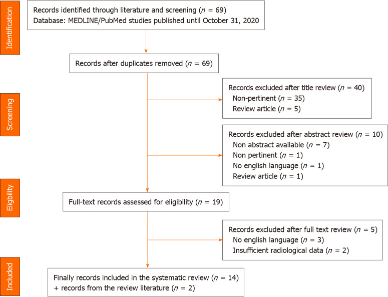
We used pertinent keywords on PubMed to select relevant studies; the articles we considered were published until October 30, 2020. Through this selection, 69 studies were identified, and 16 were finally included in the review.
Summarizing the included works' findings, we identified well-defined stages in the short follow-up time frame. A radiographic deterioration reaching a peak roughly within the first 2 wk; after the peak, an absorption process and repairing signs are observed. At later radiological follow-up, with the limitation of little evidence available, the lesions usually did not recover completely.
Following computed tomography scan evolution over time could help physicians better understand the clinical impact of COVID-19 pneumonia and manage the possible sequelae; a longer follow-up is advisable to verify the complete resolution or the presence of long-term damage.
肺炎是2019冠状病毒病(COVID-19)感染的主要表现。推荐采用胸部计算机断层扫描对该病进行初步评估;该技术也有助于监测疾病进展和评估治疗效果。
回顾目前关于使用计算机断层扫描对COVID-19相关肺部改变进行影像学随访的文献,描述COVID-19肺炎动态演变的证据,并验证影像学随访的潜在用途。
我们在PubMed上使用相关关键词筛选相关研究;纳入的文章截至2020年10月30日发表。通过此次筛选,共识别出69项研究,最终16项被纳入本综述。
总结纳入研究的结果,我们在短期随访时间框架内确定了明确的阶段。影像学恶化在大约前2周内达到高峰;高峰过后,观察到吸收过程和修复迹象。在后期影像学随访中,由于可用证据有限,病变通常未完全恢复。
随时间推移跟踪计算机断层扫描的演变情况有助于医生更好地了解COVID-19肺炎的临床影响并处理可能的后遗症;建议进行更长时间的随访以确认病变是否完全消退或是否存在长期损害。