Division of Gastroenterology, Department of Internal Medicine, Showa University Fujigaoka Hospital, 1-30 Fujigaoka, Aoba-ku, Yokohama-shi, Kanagawa, 227-8501, Japan.
Clin J Gastroenterol. 2022 Feb;15(1):210-215. doi: 10.1007/s12328-021-01532-1. Epub 2021 Oct 12.
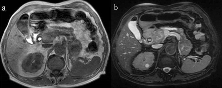
We present the case of an 86-year-old man who had undergone left nephrectomy for renal cell carcinoma (clear cell carcinoma) 22 years ago. He visited the emergency department complaining of right hypochondrial pain and fever. He was eventually diagnosed with acute cholangitis. Abdominal contrast-enhanced computed tomography showed multiple tumors in the pancreas. The tumor in the pancreatic head obstructed the distal bile duct. Endoscopic retrograde cholangiopancreatography detected bloody bile juice flowing from the papilla of Vater. Therefore, he was diagnosed with hemobilia. Cholangiography showed extrinsic compression of the distal bile duct; a 6 Fr endoscopic nasobiliary drainage tube was placed. Endoscopic ultrasound showed that the pancreas contained multiple well-defined hypoechoic masses. Endoscopic ultrasound-guided fine-needle aspiration was performed using a 22 G needle. Pathological examination revealed clear cell carcinoma, and the final diagnosis was pancreatic metastasis of renal cell carcinoma (RCC) causing hemobilia. A partially covered metallic stent was placed in the distal bile duct. Consequently, hemobilia and cholangitis were resolved.
我们报告了一例 86 岁男性病例,他 22 年前因肾细胞癌(透明细胞癌)行左肾切除术。他因右季肋部疼痛和发热就诊于急诊科。最终被诊断为急性胆管炎。腹部增强 CT 显示胰腺内有多发性肿瘤。胰头部的肿瘤阻塞了远端胆管。内镜逆行胰胆管造影(ERCP)发现从 Vater 乳头流出血性胆汁。因此,他被诊断为胆血症。胆管造影显示远端胆管受到外压性狭窄;放置了一根 6Fr 的鼻胆管引流管。内镜超声显示胰腺内有多个边界清晰的低回声肿块。使用 22G 针进行了内镜超声引导下细针抽吸。病理检查显示为透明细胞癌,最终诊断为肾细胞癌(RCC)胰腺转移导致胆血症。在远端胆管内放置了一枚部分覆膜的金属支架。随后,胆血症和胆管炎得到解决。