• 文献检索
  • 文档翻译
  • 深度研究
  • 学术资讯
  • Suppr Zotero 插件Zotero 插件
  • 邀请有礼
  • 套餐&价格
  • 历史记录
应用&插件
Suppr Zotero 插件Zotero 插件浏览器插件Mac 客户端Windows 客户端微信小程序
定价
高级版会员购买积分包购买API积分包
服务
文献检索文档翻译深度研究API 文档MCP 服务
关于我们
关于 Suppr公司介绍联系我们用户协议隐私条款
关注我们

Suppr 超能文献

核心技术专利:CN118964589B侵权必究
粤ICP备2023148730 号-1Suppr @ 2026

文献检索

告别复杂PubMed语法,用中文像聊天一样搜索,搜遍4000万医学文献。AI智能推荐,让科研检索更轻松。

立即免费搜索

文件翻译

保留排版,准确专业,支持PDF/Word/PPT等文件格式,支持 12+语言互译。

免费翻译文档

深度研究

AI帮你快速写综述,25分钟生成高质量综述,智能提取关键信息,辅助科研写作。

立即免费体验

从胸部 X 光图像中深度挖掘肺癌恶性肿瘤模型。

Deep Mining Generation of Lung Cancer Malignancy Models from Chest X-ray Images.

机构信息

Centre for Advanced Modelling and Geospatial Information Systems (CAMGIS), Faculty of Engineering and IT, University of Technology Sydney, Sydney, NSW 2007, Australia.

IBM Australia Ltd., Sydney, NSW 2000, Australia.

出版信息

Sensors (Basel). 2021 Oct 7;21(19):6655. doi: 10.3390/s21196655.

DOI:10.3390/s21196655
PMID:34640976
原文链接:https://pmc.ncbi.nlm.nih.gov/articles/PMC8513105/
Abstract

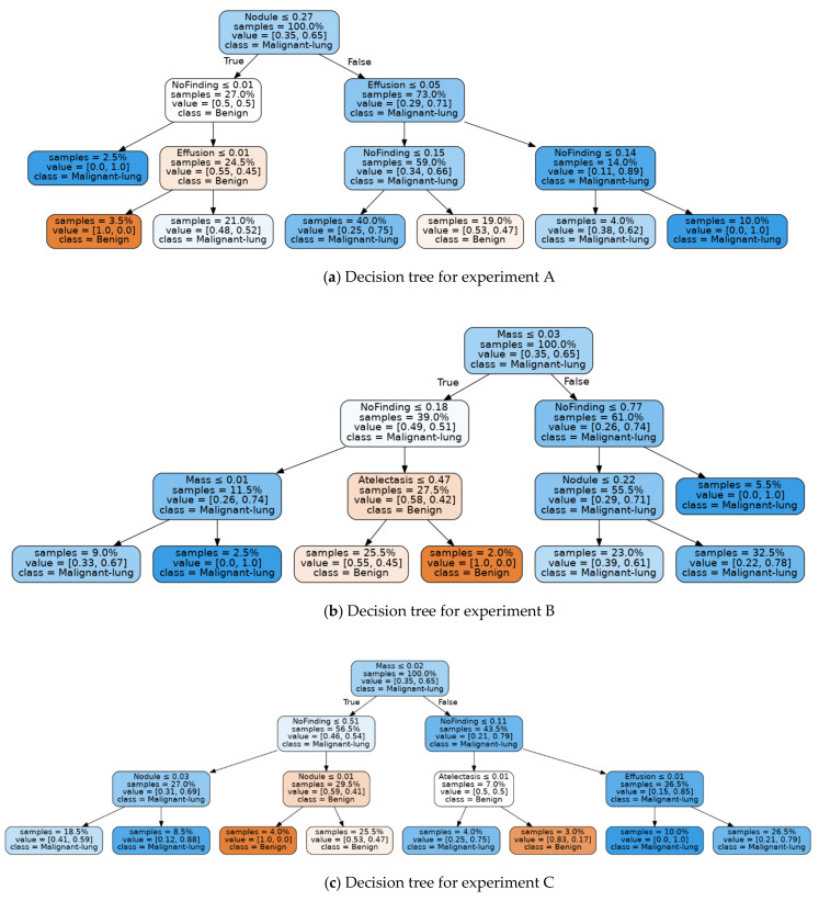
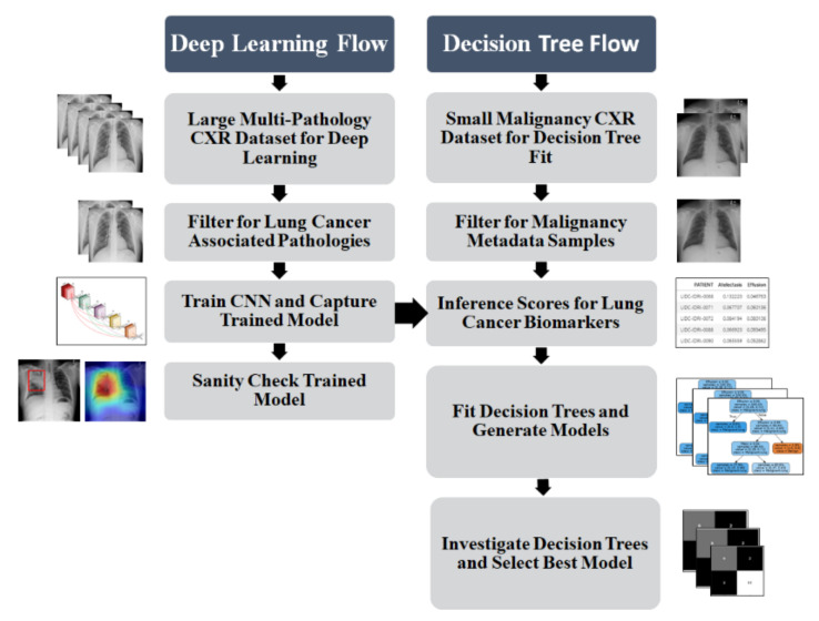
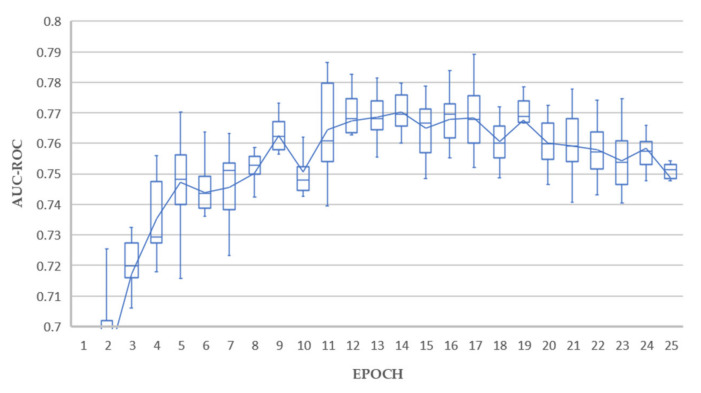
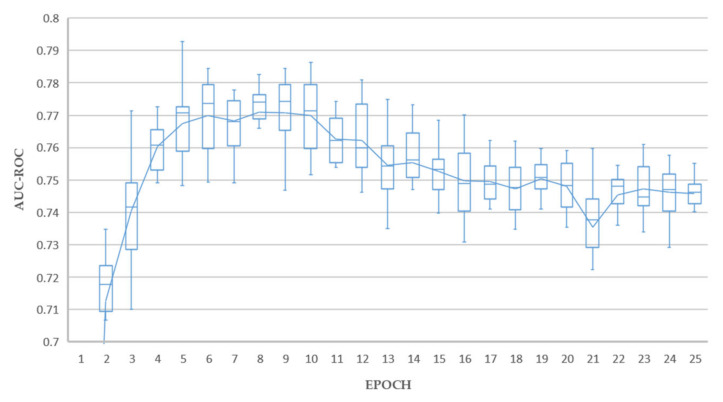
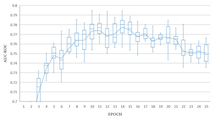
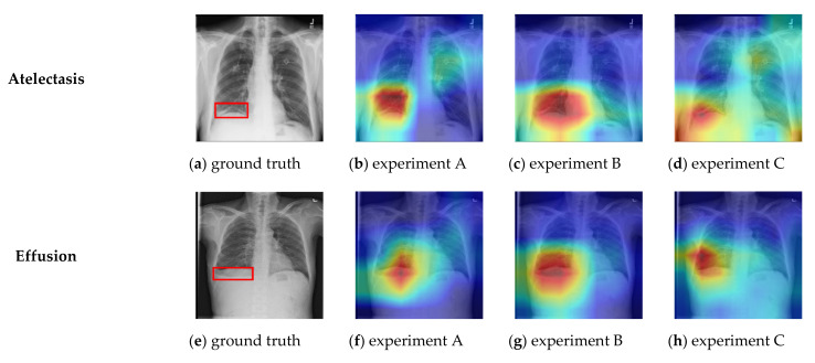
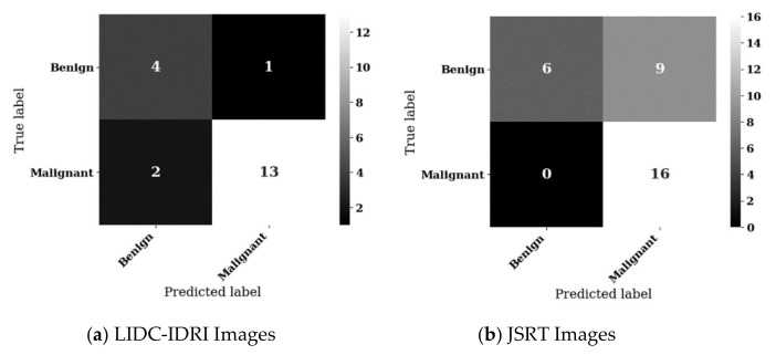
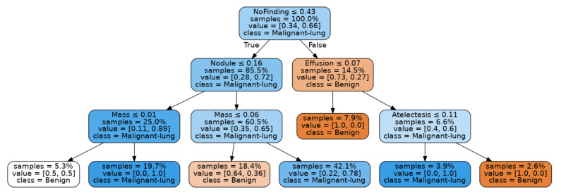
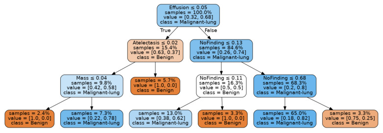
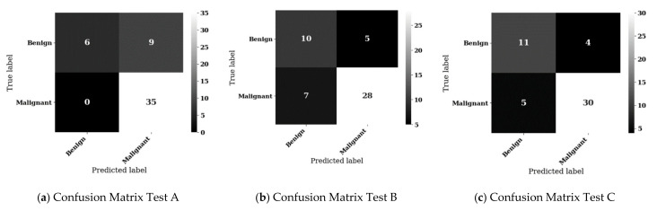
Lung cancer is the leading cause of cancer death and morbidity worldwide. Many studies have shown machine learning models to be effective in detecting lung nodules from chest X-ray images. However, these techniques have yet to be embraced by the medical community due to several practical, ethical, and regulatory constraints stemming from the "black-box" nature of deep learning models. Additionally, most lung nodules visible on chest X-rays are benign; therefore, the narrow task of computer vision-based lung nodule detection cannot be equated to automated lung cancer detection. Addressing both concerns, this study introduces a novel hybrid deep learning and decision tree-based computer vision model, which presents lung cancer malignancy predictions as interpretable decision trees. The deep learning component of this process is trained using a large publicly available dataset on pathological biomarkers associated with lung cancer. These models are then used to inference biomarker scores for chest X-ray images from two independent data sets, for which malignancy metadata is available. Next, multi-variate predictive models were mined by fitting shallow decision trees to the malignancy stratified datasets and interrogating a range of metrics to determine the best model. The best decision tree model achieved sensitivity and specificity of 86.7% and 80.0%, respectively, with a positive predictive value of 92.9%. Decision trees mined using this method may be considered as a starting point for refinement into clinically useful multi-variate lung cancer malignancy models for implementation as a workflow augmentation tool to improve the efficiency of human radiologists.

摘要

肺癌是全球癌症死亡和发病的主要原因。许多研究表明,机器学习模型在从胸部 X 光图像中检测肺结节方面非常有效。然而,由于深度学习模型的“黑盒”性质所带来的一些实际、伦理和监管方面的限制,这些技术尚未被医学界所接受。此外,胸部 X 光上可见的大多数肺结节都是良性的;因此,基于计算机视觉的肺结节检测的狭隘任务不能等同于自动肺癌检测。针对这两个问题,本研究提出了一种新颖的基于混合深度学习和决策树的计算机视觉模型,该模型将肺癌恶性程度预测表示为可解释的决策树。这个过程的深度学习部分是使用与肺癌相关的病理生物标志物的大型公共数据集进行训练的。然后,使用这些模型对来自两个独立数据集的胸部 X 光图像进行推断生物标志物评分,这些数据集都有恶性肿瘤元数据。接下来,通过对分层数据集拟合浅层决策树,并对一系列指标进行查询,挖掘多变量预测模型,以确定最佳模型。最佳决策树模型的敏感性和特异性分别为 86.7%和 80.0%,阳性预测值为 92.9%。使用这种方法挖掘的决策树可以被认为是一个起点,用于进一步细化为临床有用的多变量肺癌恶性肿瘤模型,作为工作流程增强工具,以提高人类放射科医生的工作效率。

https://cdn.ncbi.nlm.nih.gov/pmc/blobs/9a8d/8513105/b337ea874182/sensors-21-06655-g013.jpg
https://cdn.ncbi.nlm.nih.gov/pmc/blobs/9a8d/8513105/6c54eeb4974d/sensors-21-06655-g001.jpg
https://cdn.ncbi.nlm.nih.gov/pmc/blobs/9a8d/8513105/863292539e64/sensors-21-06655-g002.jpg
https://cdn.ncbi.nlm.nih.gov/pmc/blobs/9a8d/8513105/52d239faab3c/sensors-21-06655-g003a.jpg
https://cdn.ncbi.nlm.nih.gov/pmc/blobs/9a8d/8513105/5aefc3b4e078/sensors-21-06655-g004a.jpg
https://cdn.ncbi.nlm.nih.gov/pmc/blobs/9a8d/8513105/bf7e6c27cc0b/sensors-21-06655-g005.jpg
https://cdn.ncbi.nlm.nih.gov/pmc/blobs/9a8d/8513105/6f59cea43dd2/sensors-21-06655-g006.jpg
https://cdn.ncbi.nlm.nih.gov/pmc/blobs/9a8d/8513105/4cedbe22e44c/sensors-21-06655-g007.jpg
https://cdn.ncbi.nlm.nih.gov/pmc/blobs/9a8d/8513105/cf648a1696ca/sensors-21-06655-g008a.jpg
https://cdn.ncbi.nlm.nih.gov/pmc/blobs/9a8d/8513105/9c81b8a93bed/sensors-21-06655-g009.jpg
https://cdn.ncbi.nlm.nih.gov/pmc/blobs/9a8d/8513105/b0f5e78c2019/sensors-21-06655-g010.jpg
https://cdn.ncbi.nlm.nih.gov/pmc/blobs/9a8d/8513105/aabe35e916e9/sensors-21-06655-g011.jpg
https://cdn.ncbi.nlm.nih.gov/pmc/blobs/9a8d/8513105/b652bad67852/sensors-21-06655-g012.jpg
https://cdn.ncbi.nlm.nih.gov/pmc/blobs/9a8d/8513105/b337ea874182/sensors-21-06655-g013.jpg

相似文献

1
Deep Mining Generation of Lung Cancer Malignancy Models from Chest X-ray Images.从胸部 X 光图像中深度挖掘肺癌恶性肿瘤模型。
Sensors (Basel). 2021 Oct 7;21(19):6655. doi: 10.3390/s21196655.
2
Chest x-ray automated triage: A semiologic approach designed for clinical implementation, exploiting different types of labels through a combination of four Deep Learning architectures.胸部X光自动分诊:一种为临床应用设计的符号学方法,通过四种深度学习架构的组合利用不同类型的标签。
Comput Methods Programs Biomed. 2021 Jul;206:106130. doi: 10.1016/j.cmpb.2021.106130. Epub 2021 May 2.
3
Validation of a Deep Learning Algorithm for the Detection of Malignant Pulmonary Nodules in Chest Radiographs.深度学习算法在胸部 X 光片中检测恶性肺结节的验证。
JAMA Netw Open. 2020 Sep 1;3(9):e2017135. doi: 10.1001/jamanetworkopen.2020.17135.
4
Development of Debiasing Technique for Lung Nodule Chest X-ray Datasets to Generalize Deep Learning Models.开发用于肺部结节 X 射线数据集的去偏技术,以推广深度学习模型。
Sensors (Basel). 2023 Jul 21;23(14):6585. doi: 10.3390/s23146585.
5
Towards AI-augmented radiology education: a web-based application for perception training in chest X-ray nodule detection.迈向人工智能增强型放射学教育:一种用于胸部 X 射线结节检测感知训练的基于网络的应用程序。
Br J Radiol. 2023 Dec;96(1152):20230299. doi: 10.1259/bjr.20230299. Epub 2023 Oct 24.
6
Automatic creation of annotations for chest radiographs based on the positional information extracted from radiographic image reports.基于从放射影像报告中提取的位置信息,为胸部 X 光片自动创建注释。
Comput Methods Programs Biomed. 2021 Sep;209:106331. doi: 10.1016/j.cmpb.2021.106331. Epub 2021 Aug 4.
7
Automated Lung Nodule Detection and Classification Using Deep Learning Combined with Multiple Strategies.基于深度学习结合多种策略的肺结节自动检测与分类
Sensors (Basel). 2019 Aug 28;19(17):3722. doi: 10.3390/s19173722.
8
Deep learning, reusable and problem-based architectures for detection of consolidation on chest X-ray images.深度学习,可复用且基于问题的架构,用于检测胸部 X 射线图像中的实变。
Comput Methods Programs Biomed. 2020 Mar;185:105162. doi: 10.1016/j.cmpb.2019.105162. Epub 2019 Oct 31.
9
Evolving the pulmonary nodules diagnosis from classical approaches to deep learning-aided decision support: three decades' development course and future prospect.从经典方法到深度学习辅助决策支持的肺部结节诊断演进:三十年的发展历程和未来展望。
J Cancer Res Clin Oncol. 2020 Jan;146(1):153-185. doi: 10.1007/s00432-019-03098-5. Epub 2019 Nov 30.
10
AI-based improvement in lung cancer detection on chest radiographs: results of a multi-reader study in NLST dataset.基于人工智能的胸部 X 光肺癌检测改进:NLST 数据集的多读者研究结果。
Eur Radiol. 2021 Dec;31(12):9664-9674. doi: 10.1007/s00330-021-08074-7. Epub 2021 Jun 4.

引用本文的文献

1
A Thorough Review of the Clinical Applications of Artificial Intelligence in Lung Cancer.人工智能在肺癌临床应用的全面综述
Cancers (Basel). 2025 Mar 4;17(5):882. doi: 10.3390/cancers17050882.
2
Computer-assisted diagnosis for an early identification of lung cancer in chest X rays.计算机辅助诊断在胸部 X 光片中用于早期肺癌识别。
Sci Rep. 2023 May 12;13(1):7720. doi: 10.1038/s41598-023-34835-z.

本文引用的文献

1
Deep learning-enabled medical computer vision.基于深度学习的医学计算机视觉。
NPJ Digit Med. 2021 Jan 8;4(1):5. doi: 10.1038/s41746-020-00376-2.
2
A scoping review of transfer learning research on medical image analysis using ImageNet.一项关于使用ImageNet进行医学图像分析的迁移学习研究的范围综述。
Comput Biol Med. 2021 Jan;128:104115. doi: 10.1016/j.compbiomed.2020.104115. Epub 2020 Nov 13.
3
Artificial intelligence is improving the detection of lung cancer.人工智能正在提高肺癌的检测水平。
Nature. 2020 Nov;587(7834):S20-S22. doi: 10.1038/d41586-020-03157-9.
4
Triple attention learning for classification of 14 thoracic diseases using chest radiography.基于胸部 X 光的 14 种胸部疾病分类的三重注意学习。
Med Image Anal. 2021 Jan;67:101846. doi: 10.1016/j.media.2020.101846. Epub 2020 Oct 16.
5
The state of artificial intelligence-based FDA-approved medical devices and algorithms: an online database.基于人工智能且获美国食品药品监督管理局批准的医疗设备及算法的现状:一个在线数据库。
NPJ Digit Med. 2020 Sep 11;3:118. doi: 10.1038/s41746-020-00324-0. eCollection 2020.
6
Evolving the pulmonary nodules diagnosis from classical approaches to deep learning-aided decision support: three decades' development course and future prospect.从经典方法到深度学习辅助决策支持的肺部结节诊断演进:三十年的发展历程和未来展望。
J Cancer Res Clin Oncol. 2020 Jan;146(1):153-185. doi: 10.1007/s00432-019-03098-5. Epub 2019 Nov 30.
7
Multi-resolution convolutional networks for chest X-ray radiograph based lung nodule detection.基于胸部 X 射线的肺结节检测的多分辨率卷积网络。
Artif Intell Med. 2020 Mar;103:101744. doi: 10.1016/j.artmed.2019.101744. Epub 2019 Oct 28.
8
Sensitivity of chest X-ray for detecting lung cancer in people presenting with symptoms: a systematic review.胸部 X 光片检测有症状人群肺癌的敏感性:系统评价。
Br J Gen Pract. 2019 Nov 28;69(689):e827-e835. doi: 10.3399/bjgp19X706853. Print 2019 Dec.
9
Deep Learning to Assess Long-term Mortality From Chest Radiographs.深度学习评估胸部 X 光片的长期死亡率。
JAMA Netw Open. 2019 Jul 3;2(7):e197416. doi: 10.1001/jamanetworkopen.2019.7416.
10
Pleural thickening on screening chest X-rays: a single institutional study.筛查性胸部 X 光片的胸膜增厚:单机构研究。
Respir Res. 2019 Jul 5;20(1):138. doi: 10.1186/s12931-019-1116-9.