Institute for Biomedical Engineering, University of Zurich & ETH Zurich, Zurich, Switzerland.
Institute for Regenerative Medicine, University of Zurich, Zurich, Switzerland.
Mol Imaging Biol. 2022 Oct;24(5):700-709. doi: 10.1007/s11307-021-01655-4. Epub 2021 Oct 12.
Stroke is one of the most prevalent vascular diseases. Non-invasive molecular imaging methods have the potential to provide critical insights into the temporal dynamics and follow alterations of receptor expression and metabolism in ischemic stroke. The aim of this study was to assess the cannabinoid type 2 receptor (CBR) levels in transient middle cerebral artery occlusion (tMCAO) mouse models at subacute stage using positron emission tomography (PET) with our novel tracer [F]RoSMA-18-d6 and structural imaging by magnetic resonance imaging (MRI).
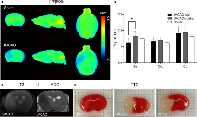
Our recently developed CBR PET tracer [F]RoSMA-18-d6 was used for imaging neuroinflammation at 24 h after reperfusion in tMCAO mice. The RNA expression levels of CBR and other inflammatory markers were analyzed by quantitative real-time polymerase chain reaction using brain tissues from tMCAO (1 h occlusion) and sham-operated mice. [F]fluorodeoxyglucose (FDG) was included for evaluation of the cerebral metabolic rate of glucose (CMRglc). In addition, diffusion-weighted imaging and T-weighted imaging were performed for anatomical reference and delineating the lesion in tMCAO mice.
mRNA expressions of inflammatory markers TNF-α, Iba1, MMP9 and GFAP, CNR2 were increased to 1.3-2.5 fold at 24 h after reperfusion in the ipsilateral compared to contralateral hemisphere of tMCAO mice, while mRNA expression of the neuronal marker MAP-2 was markedly reduced to ca. 50 %. Reduced [F]FDG uptake was observed in the ischemic striatum of tMCAO mouse brain at 24 h after reperfusion. Although higher activity of [F]RoSMA-18-d6 in ex vivo biodistribution studies and higher standard uptake value ratio (SUVR) were detected in the ischemic ipsilateral compared to contralateral striatum in tMCAO mice, the in vivo specificity of [F]RoSMA-18-d6 was confirmed only in the CBR-rich spleen.
This study revealed an increased [F]RoSMA-18-d6 measure of CBR and a reduced [F]FDG measure of CMRglc in the ischemic striatum of tMCAO mice at subacute stage. [F]RoSMA-18-d6 might be a promising PET tracer for detecting CBR alterations in animal models of neuroinflammation without neuronal loss.
中风是最常见的血管疾病之一。非侵入性分子成像方法有可能深入了解受体表达和代谢在缺血性中风中的时间动态变化和后续改变。本研究的目的是使用我们新开发的配体[F]RoSMA-18-d6 通过正电子发射断层扫描(PET)评估短暂性大脑中动脉闭塞(tMCAO)小鼠模型在亚急性阶段的大麻素 2 型受体(CBR)水平,并通过磁共振成像(MRI)进行结构成像。
我们最近开发的 CBR PET 示踪剂[F]RoSMA-18-d6 用于在 tMCAO 小鼠再灌注后 24 小时评估神经炎症。使用 tMCAO(1 小时闭塞)和假手术小鼠的脑组织通过定量实时聚合酶链反应分析 CBR 和其他炎症标志物的 RNA 表达水平。[F]氟脱氧葡萄糖(FDG)用于评估脑葡萄糖代谢率(CMRglc)。此外,还进行了弥散加权成像和 T 加权成像,以进行解剖参考并描绘 tMCAO 小鼠的病变。
与 tMCAO 小鼠对侧半球相比,再灌注后 24 小时,同侧半球中 TNF-α、Iba1、MMP9 和 GFAP 的炎症标志物的 mRNA 表达增加了 1.3-2.5 倍,而神经元标志物 MAP-2 的 mRNA 表达明显减少至约 50%。在 tMCAO 小鼠脑再灌注后 24 小时,缺血纹状体的[F]FDG 摄取减少。尽管在 tMCAO 小鼠中,在离体生物分布研究中观察到更高的[F]RoSMA-18-d6 活性和更高的标准摄取值比(SUVR),但仅在富含 CBR 的脾脏中证实了[F]RoSMA-18-d6 的体内特异性。
本研究揭示了 tMCAO 小鼠亚急性阶段缺血纹状体中 CBR 的[F]RoSMA-18-d6 测量增加和 CMRglc 的[F]FDG 测量减少。[F]RoSMA-18-d6 可能是一种有前途的 PET 示踪剂,可用于检测神经炎症动物模型中 CBR 的改变,而不会导致神经元丢失。