Institute for Biomedical Engineering, University of Zurich & ETH Zurich, Wolfgang-Pauli-Strasse 27, HIT E42.1, 8093, Zurich, Switzerland.
Zurich Neuroscience Center (ZNZ), Zurich, Switzerland.
Eur J Nucl Med Mol Imaging. 2021 Dec;48(13):4152-4170. doi: 10.1007/s00259-021-05207-4. Epub 2021 Feb 16.
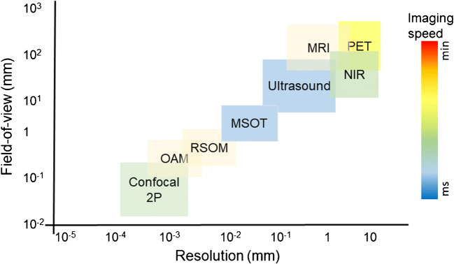
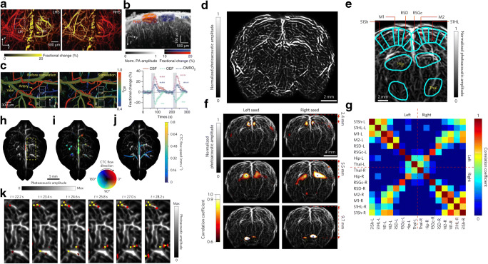
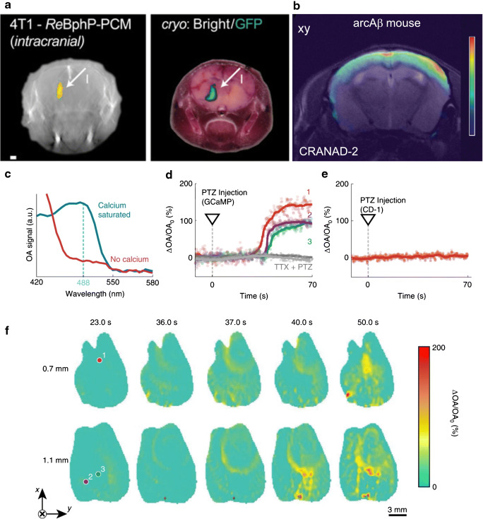
The ability to non-invasively visualize endogenous chromophores and exogenous probes and sensors across the entire rodent brain with the high spatial and temporal resolution has empowered optoacoustic imaging modalities with unprecedented capacities for interrogating the brain under physiological and diseased conditions. This has rapidly transformed optoacoustic microscopy (OAM) and multi-spectral optoacoustic tomography (MSOT) into emerging research tools to study animal models of brain diseases. In this review, we describe the principles of optoacoustic imaging and showcase recent technical advances that enable high-resolution real-time brain observations in preclinical models. In addition, advanced molecular probe designs allow for efficient visualization of pathophysiological processes playing a central role in a variety of neurodegenerative diseases, brain tumors, and stroke. We describe outstanding challenges in optoacoustic imaging methodologies and propose a future outlook.
利用光声成像模式,能够以前所未有的能力在生理和疾病状态下对大脑进行探测,该模式可无创可视化内源性发色团和外源性探针及传感器,遍及整个啮齿动物大脑,且具有高空间和时间分辨率。这使得光声显微镜(OAM)和多光谱光声断层扫描(MSOT)迅速成为研究大脑疾病动物模型的新兴研究工具。在这篇综述中,我们描述了光声成像的原理,并展示了最新的技术进展,这些进展使我们能够在临床前模型中进行高分辨率实时大脑观察。此外,先进的分子探针设计允许有效地可视化在各种神经退行性疾病、脑肿瘤和中风中起核心作用的病理生理过程。我们描述了光声成像方法学中的突出挑战,并提出了未来展望。