Departamento de Oncologia Clinica, A.C. Camargo Cancer Center, Sao Paulo, SP, BR.
Centro Internacional de Pesquisa, A.C. Camargo Cancer Center, Sao Paulo, SP, BR.
Clinics (Sao Paulo). 2021 Oct 11;76:e2971. doi: 10.6061/clinics/2021/e2971. eCollection 2021.
Breast cancer (BC) is the most common neoplasm in women. Biopsy of metastatic lesions is recommended to confirm estrogen receptor (ER), progesterone receptor (PR), and human epidermal growth factor receptor 2 (HER2) status as there are discrepancies in these patterns between primary tumors and metastases in up to 40% of the cases. Circulating tumor cells (CTCs) are related to BC outcomes and could potentially be an alternative to the invasive procedures of metastasis rebiopsy. ISET® technology is not currently employed to detect CTCs in patients with BC. Emerging data support that the characterization of CTC protein expression can refine its prognostic value. Transforming growth factor (TGF)-β plays a role in BC progression and invasiveness. Thus, in this study, we aimed to compare ER, PR, and HER2 expression in primary tumors, CTCs, and metastases and evaluate TGF-β type 1 receptor (TGF-β RI) expression in CTCs as prognostic factor for progression free survival (PFS) and overall survival (OS).
This prospective study was conducted at the A.C. Camargo Cancer Center, Brazil. Blood samples were processed in ISET® (Isolation by SizE of Tumors, Rarecells, France) before computed tomography-guided biopsy of suspected metastatic lesions. Protein expression levels in CTCs were compared to those in primary tumors/metastases (medical records).
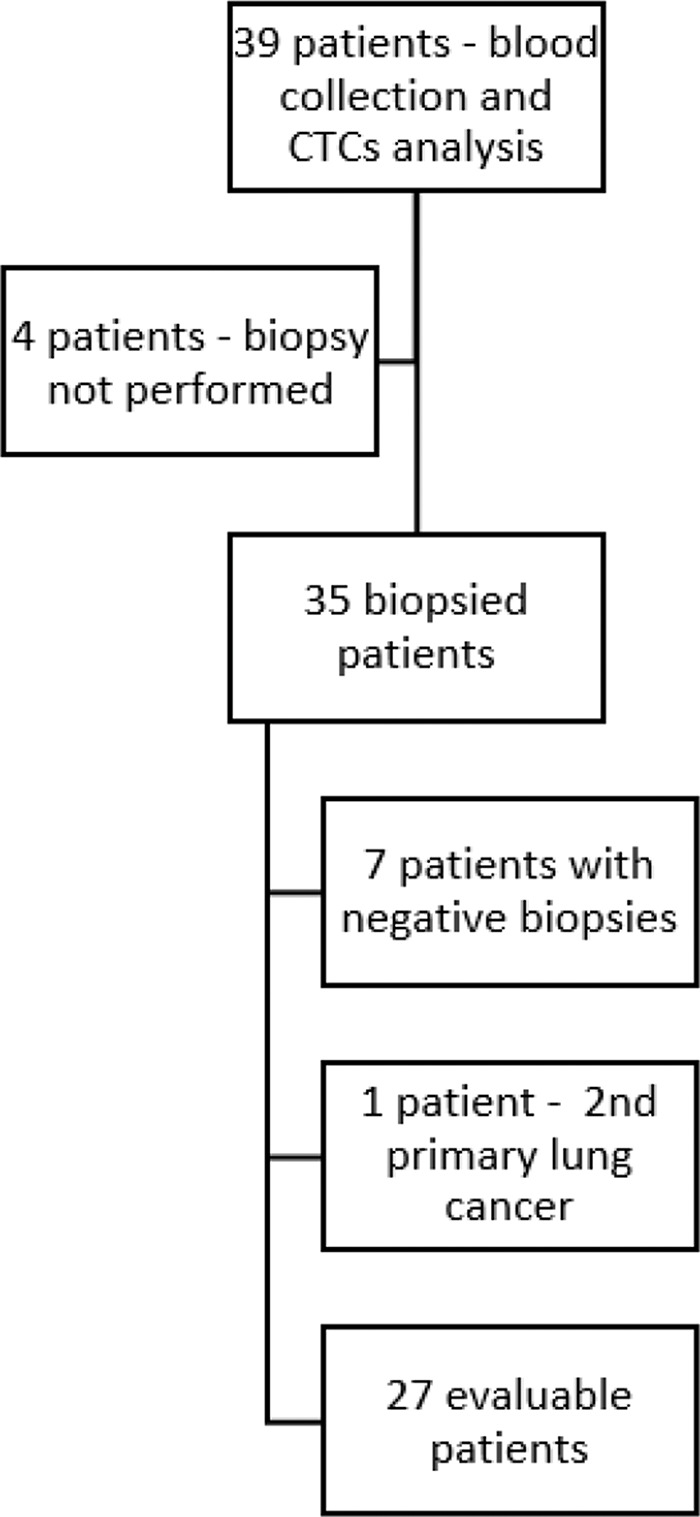
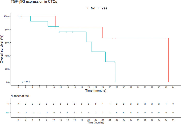
Of the 39 patients initially included, 27 underwent both biopsies of metastases and blood collection and were considered for analysis. The concordance rates for ER, PR, and HER2 expression between primary tumors and metastases were high. No loss of HER2 expression at any metastasis site and retention of the same pattern of protein expression in all triple-negative (TN) tumors (92.5%, 81.5% and 96.2% respectively) (p<0.0001) was observed. When metastases/CTCs were classified as TN/non-TN, CTCs showed high specificity (93%), accuracy (84.2%), and negative predictive value (88%). The median OS of patients without TGF-β RI expression in CTCs was 42.6 versus 20.8 months for TGF-β RI expression-positive ones (p>0.05).
The role of CTCs detected by ISET has not yet been established in BC. Here, we suggest that this methodology may be useful to evaluate metastasis in non-TN cases as well as TGF-β RI expression in CTCs, which may impact patient survival. Due to sample limitations, future studies must focus on specific BC subtypes and an expansion of the cohort.
乳腺癌(BC)是女性中最常见的肿瘤。由于原发性肿瘤和转移灶之间存在高达 40%的不一致,因此建议对转移性病变进行活检以确认雌激素受体(ER)、孕激素受体(PR)和人表皮生长因子受体 2(HER2)状态,以确定转移灶的这些模式。循环肿瘤细胞(CTC)与 BC 结果相关,并且可能是对转移再活检的侵入性程序的替代方法。目前尚未使用 ISET®技术检测 BC 患者的 CTC。新兴数据支持 CTC 蛋白表达的特征可以细化其预后价值。转化生长因子(TGF)-β 在 BC 的进展和侵袭中起作用。因此,在这项研究中,我们旨在比较原发性肿瘤、CTC 和转移灶中 ER、PR 和 HER2 的表达,并评估 CTC 中 TGF-β Ⅰ型受体(TGF-β RI)的表达作为无进展生存期(PFS)和总生存期(OS)的预后因素。
这项前瞻性研究在巴西 A.C. Camargo 癌症中心进行。在对疑似转移性病变进行计算机断层扫描引导活检之前,通过 ISET®(肿瘤大小分离,稀有细胞,法国)处理血液样本。将 CTC 中的蛋白表达水平与原发性肿瘤/转移灶(病历)进行比较。
最初纳入的 39 名患者中,有 27 名患者同时进行了转移灶活检和血液采集,并进行了分析。原发性肿瘤和转移灶之间 ER、PR 和 HER2 表达的一致性率较高。未观察到任何转移部位的 HER2 表达缺失,所有三阴性(TN)肿瘤的蛋白表达模式保持一致(分别为 92.5%、81.5%和 96.2%)(p<0.0001)。当将转移灶/CTC 分为 TN/非 TN 时,CTC 表现出高特异性(93%)、准确性(84.2%)和阴性预测值(88%)。在 CTC 中无 TGF-β RI 表达的患者中位 OS 为 42.6 个月,而 TGF-β RI 表达阳性的患者为 20.8 个月(p>0.05)。
ISET 检测的 CTC 在 BC 中的作用尚未确定。在这里,我们建议该方法可能有助于评估非 TN 病例的转移情况,以及 CTC 中的 TGF-β RI 表达,这可能会影响患者的生存。由于样本限制,未来的研究必须侧重于特定的 BC 亚型和扩大队列。