Naganawa Shinji, Ito Rintaro, Kawamura Mariko, Taoka Toshiaki, Yoshida Tadao, Sone Michihiko
Department of Radiology, Nagoya University Graduate School of Medicine.
Department of Otorhinolaryngology, Nagoya University Graduate School of Medicine.
Magn Reson Med Sci. 2023 Jan 1;22(1):45-55. doi: 10.2463/mrms.mp.2021-0100. Epub 2021 Oct 16.
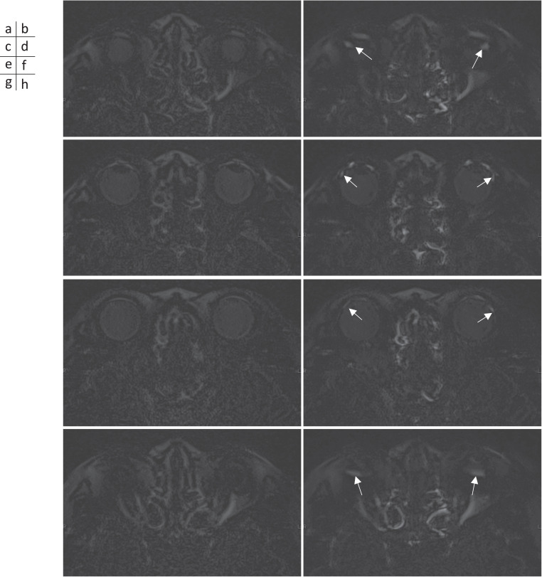
Peripheral retinal leakage (PRL) of contrast medium from the ora serrata (i.e., the peripheral part of the retina) was recently reported in normal eyes using ultra-widefield fluorescein angiography. We occasionally see PRL of gadolinium-based contrast agents (GBCAs) in the vitreous from the temporal and inferior sides of the ora serrata on MR images of subjects without ophthalmic disease. In this study, we retrospectively evaluated these MR images to determine if PRL was associated with aging. We also evaluated whether the initial leakage appeared in the temporal and inferior sides, and whether there was uniform distribution within the vitreous after 24 hours.
In 127 subjects (9 volunteers, 85 patients with sudden deafness, and 33 patients with a suspicion of endolymphatic hydrops), pre- and post-contrast-enhanced heavily T2-weighted 3D-fluid attenuated inversion recovery (FLAIR) images were obtained. The presence or absence of PRL was subjectively evaluated. For patients with a suspicion of endolymphatic hydrops, 3D-real inversion recovery (IR) images were also obtained at pre-, 10 mins, 4 hours, and 24 hours after intravenous administration (IV) of GBCA. Four circular ROIs were placed in the vitreous humor and the signal intensity was measured.
In the cases with PRL (n = 88) and without PRL (n = 47), the median age was 59 and 47 years, respectively (P = 0.001). At 4 hours after IV-GBCA, the mean signal increase in the inferior temporal ROI was greater than all the other ROIs. At 24 hours after IV-GBCA, no significant difference in signal intensity was observed for the four ROIs.
PRL of GBCA is age-dependent and occurs mainly from the inferior temporal side of the ora serrata. The contrast effect was uniformly distributed at 24 hours after IV-GBCA. Future observations in a variety of diseases will determine the clinical significance of these findings.
最近有报道称,使用超广角荧光素血管造影术在正常眼睛中观察到造影剂从锯齿缘(即视网膜的周边部分)发生周边视网膜渗漏(PRL)。在无眼科疾病受试者的磁共振成像(MRI)上,我们偶尔会看到基于钆的造影剂(GBCA)从锯齿缘的颞侧和下方进入玻璃体导致的PRL。在本研究中,我们对这些MRI图像进行回顾性评估,以确定PRL是否与年龄相关。我们还评估了最初的渗漏是否出现在颞侧和下方,以及24小时后玻璃体中的分布是否均匀。
对127名受试者(9名志愿者、85名突发性耳聋患者和33名疑似内淋巴积水患者)进行了对比增强前后的重T2加权三维液体衰减反转恢复(FLAIR)成像。对PRL的有无进行主观评估。对于疑似内淋巴积水的患者,在静脉注射GBCA前、注射后10分钟、4小时和24小时还获取了三维真实反转恢复(IR)图像。在玻璃体内放置四个圆形感兴趣区(ROI)并测量信号强度。
有PRL的病例(n = 88)和无PRL的病例(n = 47),中位年龄分别为59岁和47岁(P = 0.001)。静脉注射GBCA后4小时,颞下ROI的平均信号增加大于所有其他ROI。静脉注射GBCA后24小时,四个ROI的信号强度未观察到显著差异。
GBCA的PRL与年龄相关,主要发生在锯齿缘的颞下侧。静脉注射GBCA后24小时,对比效果均匀分布。未来对各种疾病的观察将确定这些发现的临床意义。