GRN Hospital Weinheim, Department of Cardiology, Vascular Medicine and Pneumology, Weinheim, Germany.
Cardiac Imaging Center Weinheim, Hector Foundation, Weinheim, Germany.
Vasc Health Risk Manag. 2021 Oct 23;17:661-673. doi: 10.2147/VHRM.S295376. eCollection 2021.
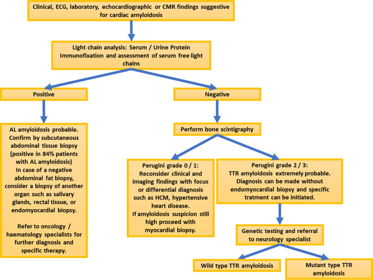
Among non-ischemic cardiomyopathies, cardiac amyloidosis is one of the most common, being caused by extracellular depositions of amyloid fibrils in the myocardium. Two main forms of cardiac amyloidosis are known so far, including 1) light-chain (AL) amyloidosis caused by monoclonal production of light-chains, and 2) transthyretin (ATTR) amyloidosis, caused by dissociation of the transthyretin tetramer into monomers. Both AL and ATTR amyloidosis are progressive diseases with median survival from diagnosis of less than 6 months and 3 to 5 years, respectively, if untreated. In this regard, death occurs in most patients due to cardiac causes, mainly congestive heart failure, which can be prevented due to the presence of effective, life-saving treatment regimens. Therefore, early diagnosis of cardiac amyloidosis is crucial more than ever. However, diagnosis of cardiac amyloidosis may be challenging due to variable clinical manifestations and the perceived rarity of the disease. In this regard, clinical and laboratory reg flags are available, which may help clinicians to raise suspicion of cardiac amyloidosis. In addition, advances in cardiovascular imaging have already revealed a higher prevalence of cardiac amyloidosis in specific populations, so that the diagnosis especially of ATTR amyloidosis has experienced a >30-fold increase during the past ten years. The goal of our review article is to summarize these findings and provide a practical approach for clinicians on how to use cardiovascular imaging techniques, such as echocardiography, cardiac magnetic resonance, bone scintigraphy and, if required, organ biopsy within predefined diagnostic algorithms for the diagnostic work-up of patients with suspected cardiac amyloidosis. In addition, two clinical cases and practical tips are provided in this context.
在非缺血性心肌病中,心脏淀粉样变性是最常见的疾病之一,其病因是心肌细胞外沉积淀粉样纤维。目前已知有两种主要形式的心脏淀粉样变性,包括 1)由单克隆轻链产生引起的轻链(AL)淀粉样变性,和 2)由转甲状腺素蛋白(ATTR)四聚体解聚为单体引起的转甲状腺素蛋白(ATTR)淀粉样变性。AL 和 ATTR 淀粉样变性都是进行性疾病,如果未经治疗,从中位数诊断到存活的时间分别不到 6 个月和 3 至 5 年。在这方面,大多数患者因心脏原因死亡,主要是充血性心力衰竭,如果有有效的、救命的治疗方案,这种情况是可以预防的。因此,及早诊断心脏淀粉样变性比以往任何时候都更为重要。然而,由于临床表现的多变性和对该病的罕见性的认知,心脏淀粉样变性的诊断可能具有挑战性。在这方面,临床和实验室检查标志可用,这可能有助于临床医生怀疑心脏淀粉样变性。此外,心血管成像技术的进步已经揭示了在特定人群中心脏淀粉样变性的患病率更高,因此,在过去十年中,特别是 ATTR 淀粉样变性的诊断增加了 30 多倍。我们的综述文章的目的是总结这些发现,并为临床医生提供实用的方法,即在疑似心脏淀粉样变性患者的诊断工作中,如何使用心血管成像技术,如超声心动图、心脏磁共振、骨闪烁扫描,如果需要,还可以使用器官活检,并在既定的诊断算法中进行。此外,在这方面还提供了两个临床病例和实用提示。