Biophotonics@Tyndall, IPIC, Tyndall National Institute, Lee Maltings, Dyke Parade, Cork, Ireland.
University College Cork, Department of Physics, Cork, Ireland.
J Biomed Opt. 2021 Nov;27(7). doi: 10.1117/1.JBO.27.7.074707.
Gas in scattering media absorption spectroscopy (GASMAS) enables noninvasive gas sensing in the body. It is developing as a tool for diagnosis and monitoring of respiratory conditions in neonates. Phantom models with relevant features to the clinical translation of GASMAS technology are necessary to understand technical challenges and potential applications of this technique. State-of-the-art phantoms designed for this purpose have focused on the optical properties and anthropomorphic geometry of the thorax, contributing to the source-detector placement, design, and optimization. Lung phantom mimicking the alveolar anatomy has not been included in the existent models due to the inherent complexity of the tissue. We present a simplified model that recreates inflated alveoli embedded in lung phantom.
The goal of this study was to build a lung model with air-filled structures mimicking inflated alveoli surrounded by optical phantom with accurate optical properties (μa = 0.50 cm - 1 and μs'=5.4 cm-1) and physiological parameters [37°C and 100% relative humidity (RH)], and to control the air volume within the phantom to demonstrate the feasibility of GASMAS in sensing changes in pulmonary air volume.
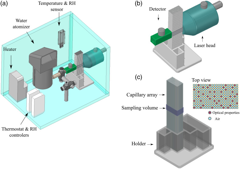
The lung model was built using a capillary structure with analogous size to alveolar units. Part of the capillaries were filled with liquid lung optical phantom to recreate scattering and absorption, whereas empty capillaries mimicked air filled alveoli. The capillary array was placed inside a custom-made chamber that maintained pulmonary temperature and RH. The geometry of the chamber permitted the placement of the laser head and detector of a GASMAS bench top system (MicroLab Dual O2 / H2O), to test the changes in volume of the lung model in transmittance geometry.
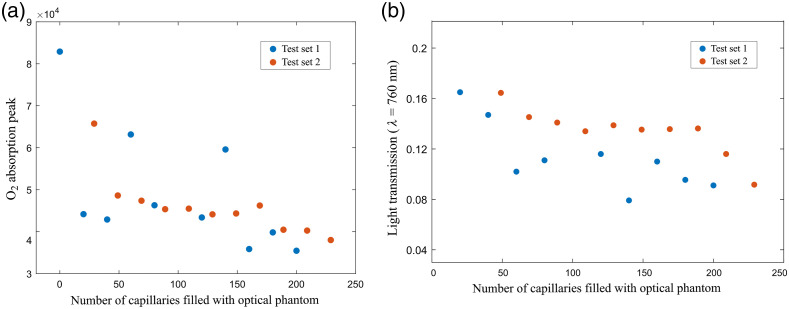
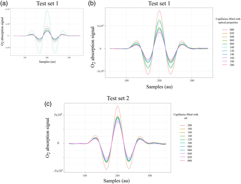
The lung tissue model with air volume range from 6.89 × 10 - 7 m3 to 1.80 × 10 - 3 m3 was built. Two measurement sets, with 10 different capillary configurations each, were arranged to increase or decrease progressively (in steps of 3.93 × 10 - 8 m3) the air volume in the lung model. The respective GASMAS data acquisition was performed for both data sets. The maximum absorption signal was obtained for configurations with the highest number of air-filled capillaries and decreased progressively when the air spaces were replaced by capillaries filled with liquid optical phantom. Further studies are necessary to define the minimum and maximum volume of air that can be measured with GASMAS-based devices for different source-detector geometries.
The optical properties and the structure of tissue from the respiratory zone have been modeled using a simplified capillary array immersed in a controlled environment chamber at pulmonary temperature and RH. The feasibility of measuring volume changes with GASMAS technique has been proven, stating a new possible application of GASMAS technology in respiratory treatment and diagnostics.
在散射介质吸收光谱(GASMAS)中的气体能够实现体内的非侵入式气体感应。它正在发展成为诊断和监测新生儿呼吸状况的工具。具有与 GASMAS 技术临床转化相关特征的幻影模型对于理解该技术的技术挑战和潜在应用是必要的。为此目的设计的最先进的幻影模型侧重于胸部的光学特性和拟人化几何形状,有助于源探测器的放置、设计和优化。由于组织的固有复杂性,尚未在现有模型中包括模拟肺泡解剖结构的肺幻影。我们提出了一种简化的模型,该模型再现了充满空气的结构,模拟了充满空气的肺泡,肺泡嵌入在肺幻影中。
本研究的目的是构建一个带有充满空气的结构的肺模型,这些结构模拟充满空气的肺泡,周围是具有准确光学特性(μa = 0.50 cm-1 和 μs'=5.4 cm-1)和生理参数[37°C 和 100%相对湿度(RH)]的光学幻影,并控制幻影内的空气量,以证明 GASMAS 在感应肺部空气量变化方面的可行性。
使用类似于肺泡单元大小的毛细管结构构建肺模型。部分毛细管充满液体肺光学幻影以再现散射和吸收,而空毛细管模拟充满空气的肺泡。毛细管阵列放置在一个定制的腔室内,该腔室保持肺部温度和 RH。腔室的几何形状允许放置 GASMAS 台式系统(MicroLab Dual O2/H2O)的激光头和探测器,以测试透射几何结构中肺模型体积的变化。
构建了空气体积范围为 6.89×10⁻⁷ m3 至 1.80×10⁻³ m3 的肺组织模型。安排了两组测量,每组有 10 种不同的毛细管配置,以逐渐增加或减少(每次增加 3.93×10⁻⁸ m3)肺模型中的空气体积。对两组数据分别进行了相应的 GASMAS 数据采集。当空气空间被充满液体光学幻影的毛细管取代时,获得了具有最高数量充满空气的毛细管的配置的最大吸收信号,并且信号逐渐降低。需要进一步的研究来确定不同源探测器几何形状下基于 GASMAS 的设备可以测量的最小和最大空气体积。
使用浸入受控环境室中的简化毛细管阵列模拟了呼吸区的光学特性和组织结构,该环境室处于肺部温度和 RH。已经证明了使用 GASMAS 技术测量体积变化的可行性,提出了 GASMAS 技术在呼吸治疗和诊断中的新的潜在应用。