Deans Michael R
Department of Surgery, Division of Otolaryngology, University of Utah School of Medicine, Salt Lake City, UT, United States.
Department of Neurobiology, University of Utah School of Medicine, Salt Lake City, UT, United States.
Front Neurosci. 2021 Oct 18;15:742391. doi: 10.3389/fnins.2021.742391. eCollection 2021.
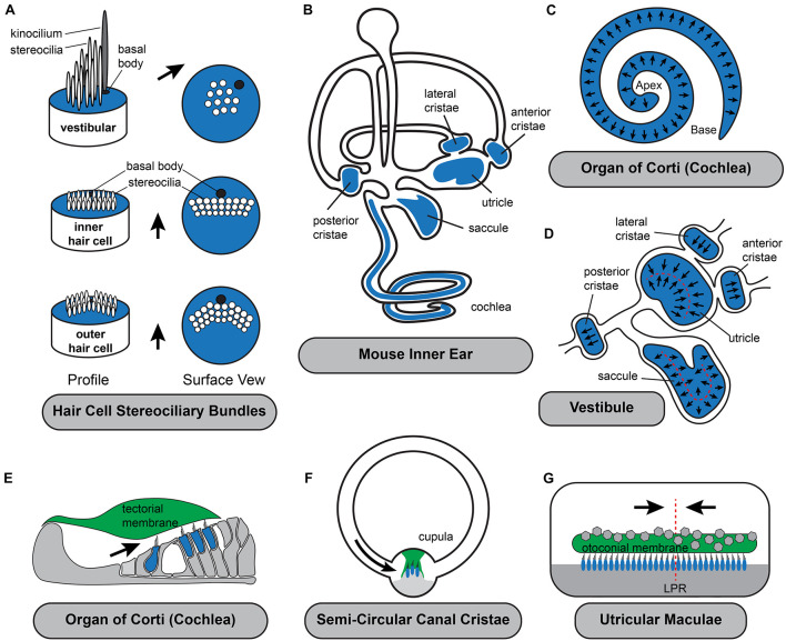
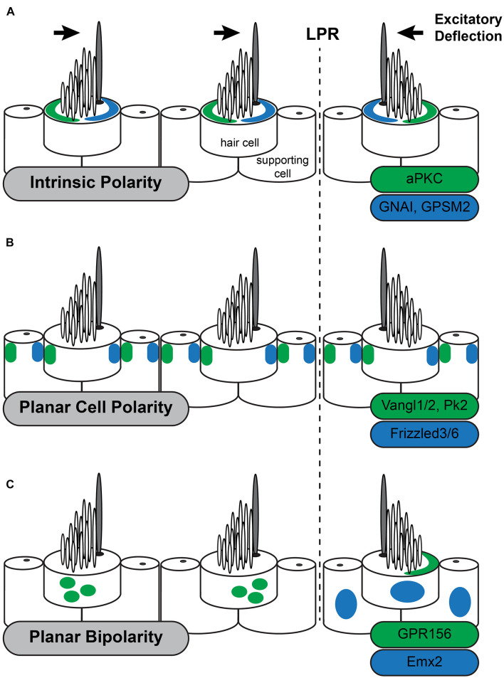
Planar polarity describes the organization and orientation of polarized cells or cellular structures within the plane of an epithelium. The sensory receptor hair cells of the vertebrate inner ear have been recognized as a preeminent vertebrate model system for studying planar polarity and its development. This is principally because planar polarity in the inner ear is structurally and molecularly apparent and therefore easy to visualize. Inner ear planar polarity is also functionally significant because hair cells are mechanosensors stimulated by sound or motion and planar polarity underlies the mechanosensory mechanism, thereby facilitating the auditory and vestibular functions of the ear. Structurally, hair cell planar polarity is evident in the organization of a polarized bundle of actin-based protrusions from the apical surface called stereocilia that is necessary for mechanosensation and when stereociliary bundle is disrupted auditory and vestibular behavioral deficits emerge. Hair cells are distributed between six sensory epithelia within the inner ear that have evolved unique patterns of planar polarity that facilitate auditory or vestibular function. Thus, specialized adaptations of planar polarity have occurred that distinguish auditory and vestibular hair cells and will be described throughout this review. There are also three levels of planar polarity organization that can be visualized within the vertebrate inner ear. These are the intrinsic polarity of individual hair cells, the planar cell polarity or coordinated orientation of cells within the epithelia, and planar bipolarity; an organization unique to a subset of vestibular hair cells in which the stereociliary bundles are oriented in opposite directions but remain aligned along a common polarity axis. The inner ear with its complement of auditory and vestibular sensory epithelia allows these levels, and the inter-relationships between them, to be studied using a single model organism. The purpose of this review is to introduce the functional significance of planar polarity in the auditory and vestibular systems and our contemporary understanding of the developmental mechanisms associated with organizing planar polarity at these three cellular levels.
平面极性描述了上皮平面内极化细胞或细胞结构的组织和方向。脊椎动物内耳的感觉受体毛细胞已被公认为研究平面极性及其发育的卓越脊椎动物模型系统。这主要是因为内耳中的平面极性在结构和分子层面都很明显,因此易于观察。内耳平面极性在功能上也很重要,因为毛细胞是由声音或运动刺激的机械传感器,平面极性是机械感觉机制的基础,从而促进耳朵的听觉和前庭功能。在结构上,毛细胞平面极性在从顶端表面伸出的基于肌动蛋白的极化束(称为静纤毛)的组织中很明显,这对于机械感觉是必需的,当静纤毛束被破坏时,听觉和前庭行为缺陷就会出现。毛细胞分布在内耳的六个感觉上皮之间,这些上皮已经进化出独特的平面极性模式,以促进听觉或前庭功能。因此,已经出现了区分听觉和前庭毛细胞的平面极性的特殊适应,本文将对此进行描述。在脊椎动物内耳中还可以观察到三个层次的平面极性组织。这些是单个毛细胞的固有极性、上皮内细胞的平面细胞极性或协调方向,以及平面双极性;这是前庭毛细胞亚群特有的一种组织,其中静纤毛束的方向相反,但仍沿共同的极性轴排列。内耳及其听觉和前庭感觉上皮的补充,使得可以使用单一模式生物来研究这些层次以及它们之间的相互关系。本综述的目的是介绍平面极性在听觉和前庭系统中的功能意义,以及我们目前对在这三个细胞水平上组织平面极性相关发育机制的理解。