Danilkowicz Richard M, Allen Nicholas B, Grimm Nate, Nettles Dana L, Nunley James A, Easley Mark E, Adams Samuel B
Department of Orthopaedic Surgery, Duke University Medical Center, Durham, North Carolina, USA.
Idaho Sports Medicine Institute, Boise, Idaho, USA.
Orthop J Sports Med. 2021 Oct 29;9(10):23259671211040535. doi: 10.1177/23259671211040535. eCollection 2021 Oct.
The most common first-line treatment of osteochondral lesions of the talus (OLTs) is microfracture. Although many patients do well with this procedure, a number fail and require reoperation. The mechanism of failure of microfracture is unknown, and to our knowledge there has been no research characterizing failed microfracture regarding histological and inflammatory makeup of these lesions that may contribute to failure.
To characterize the structural and biochemical makeup of failed microfracture lesions.
Case series; Level of evidence, 4.
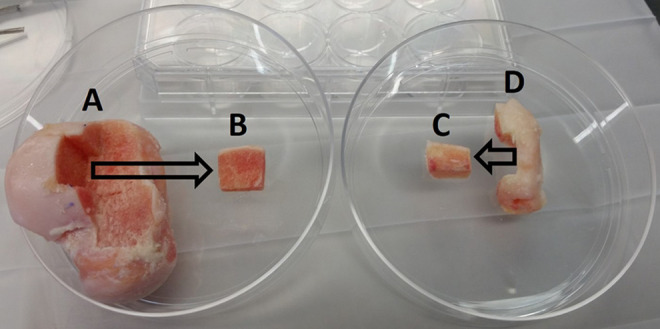
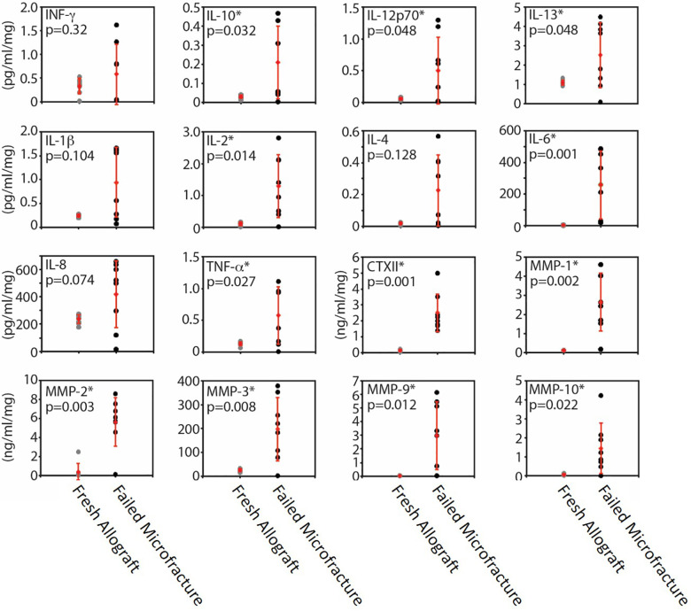
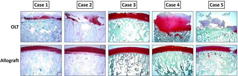
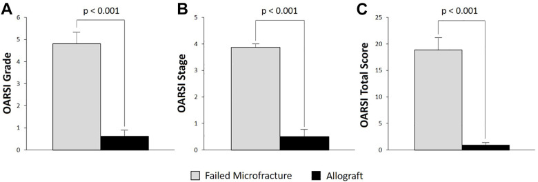
Specimens from 8 consecutive patients with symptomatic OLTs after microfracture who later underwent fresh osteochondral allograft transplantation were analyzed. For each patient, the failed microfracture specimen and a portion of the fresh allograft replacement tissue were collected. The allograft served as a control. Histology of the failed microfracture and the allograft replacement was scored using the Osteoarthritis Research Society International (OARSI) system. Surface roughness was also compared. In addition, tissue culture supernatants were analyzed for 16 secreted cytokines and matrix metalloproteinases (MMPs) responsible for inflammation, pain, cartilage damage, and chondrocyte death.
The OARSI grade, stage, and total score as well as surface smoothness were significantly worse in the failed microfracture sample, indicating better cartilage and bone morphology for the allografts compared with the failed microfracture lesions. Analyzed cytokines and MMPs were significantly elevated in the microfracture tissue culture supernatants when compared with fresh osteochondral tissue supernatants.
These data demonstrate a significantly rougher cartilage surface, cartilage and subchondral bone histology that more closely resembles osteoarthritis, and elevated inflammatory cytokines and MMPs responsible for pain, inflammation, cartilage damage, and chondrocyte death when compared with fresh osteochondral allografts used as controls.
距骨骨软骨损伤(OLTs)最常见的一线治疗方法是微骨折术。尽管许多患者接受该手术后效果良好,但仍有一些患者治疗失败,需要再次手术。微骨折术失败的机制尚不清楚,据我们所知,尚无研究对失败的微骨折术进行组织学和炎症构成特征分析,而这些特征可能导致手术失败。
对失败的微骨折术损伤的结构和生化构成进行特征分析。
病例系列研究;证据等级为4级。
对8例连续接受微骨折术后出现症状性OLTs且随后接受新鲜骨软骨同种异体移植的患者的标本进行分析。对于每位患者,收集失败的微骨折术标本和一部分新鲜同种异体移植替代组织。同种异体移植组织作为对照。使用国际骨关节炎研究学会(OARSI)系统对失败的微骨折术和同种异体移植替代组织进行组织学评分。同时比较表面粗糙度。此外,分析组织培养上清液中16种与炎症、疼痛、软骨损伤和软骨细胞死亡相关的分泌细胞因子和基质金属蛋白酶(MMPs)。
失败的微骨折术样本的OARSI分级、分期和总分以及表面光滑度明显更差,表明与失败的微骨折术损伤相比,同种异体移植组织的软骨和骨形态更好。与新鲜骨软骨组织上清液相比,微骨折术组织培养上清液中的细胞因子和MMPs分析结果明显升高。
与用作对照的新鲜骨软骨同种异体移植组织相比,这些数据表明失败的微骨折术损伤的软骨表面明显更粗糙,软骨和软骨下骨组织学更类似于骨关节炎,且与疼痛、炎症、软骨损伤和软骨细胞死亡相关的炎症细胞因子和MMPs升高。