Tapia Daisy, Floriolli David, Han Eric, Lee Grace, Paganini-Hill Annlia, Wang Stephani, Zandihaghighi Setarah, Kimonis Virginia, Fisher Mark
Division of Genetic and Genomic Medicine, Department of Pediatrics, University of California Irvine Medical Center, CA, USA.
Department of Radiological Sciences, Neuroradiology, University of California Irvine Medical Center, CA, USA.
Mol Genet Metab Rep. 2021 Oct 21;29:100815. doi: 10.1016/j.ymgmr.2021.100815. eCollection 2021 Dec.
To characterize the prevalence of brain ischemia and cerebral small vessel disease in a cohort of patients with Fabry disease (FD) seen at an academic medical center.
FD is an inherited X-linked lysosomal storage disorder with central nervous system involvement. Limited data are available in the literature on the cerebrovascular neuroimaging findings in FD, and the reported prevalence of stroke symptoms and cerebral small vessel disease has varied widely.
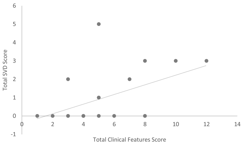
DESIGN/METHODS: Brain MRI was performed in 21 patients with FD followed at University of California Irvine Medical Center. Stroke symptoms were assessed and quantification of cerebral microvascular disease was performed using small vessel disease (SVD) score. Lacunes and deep white matter hyperintensities were scored on a four-point scale of 0 (absent) and 1-3 to account for increasing severity; microbleeds were scored 0 (absent) or 1 (present). The total SVD score is the sum of the three components and ranges from 0 to 7.
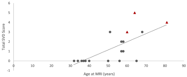
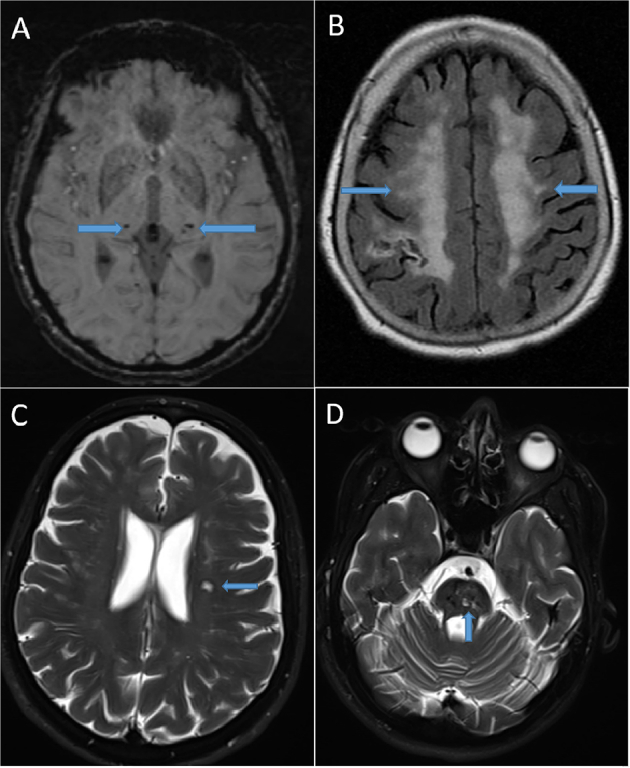
Nearly 43% (9/21) of our FD cohort (aged 32-81 years, mean = 50) had a SVD score of one or higher, all of whom were aged 50 or more years. The most common MRI-defined SVD was white matter hyperintensities (9/9, 100%), followed by microbleeds (6/9, 66%), and lacunes (3/9, 33%). The three patients with previous strokes had some of the highest SVD scores reported in the cohort (scores 3-5).
In this cohort, the prevalence of SVD (43%) was three times higher than prevalence of stroke symptoms. SVD score was highest in the those who had experienced a stroke. These findings emphasize the importance of routine MRI screening of patients with FD in order to identify and treat high risk patients.
描述在一家学术医疗中心就诊的法布里病(FD)患者队列中脑缺血和脑小血管疾病的患病率。
FD是一种遗传性X连锁溶酶体贮积症,累及中枢神经系统。关于FD脑血管神经影像学表现的文献资料有限,且报道的中风症状和脑小血管疾病患病率差异很大。
设计/方法:对加利福尼亚大学欧文医学中心随访的21例FD患者进行脑部MRI检查。评估中风症状,并使用小血管疾病(SVD)评分对脑微血管疾病进行量化。腔隙和深部白质高信号根据0(无)和1 - 3的四分制进行评分,以反映严重程度增加;微出血评分为0(无)或1(有)。总SVD评分是三个组成部分的总和,范围为0至7。
我们的FD队列中近43%(9/21)(年龄32 - 81岁,平均 = 50岁)的SVD评分为1或更高,所有这些患者年龄均在50岁及以上。MRI定义的最常见SVD是白质高信号(9/9,100%),其次是微出血(6/9,66%)和腔隙(3/9,33%)。三名既往有中风史的患者在队列中报告的SVD评分较高(评分3 - 5)。
在该队列中,SVD的患病率(43%)比中风症状的患病率高两倍。中风患者的SVD评分最高。这些发现强调了对FD患者进行常规MRI筛查以识别和治疗高危患者的重要性。