Department of Pediatrics I, Neonatology & Experimental Perinatal Neurosciences, University Hospital Essen, University Duisburg-Essen, Hufelandstr. 55, 45147, Essen, Germany.
Center for Translational Neuro-and Behavioral Sciences (C-TNBS), University Hospital Essen, University Duisburg-Essen, Essen, Germany.
J Neuroinflammation. 2021 Nov 13;18(1):266. doi: 10.1186/s12974-021-02314-9.
Neonatal encephalopathy due to hypoxia-ischemia (HI) is a leading cause of death and disability in term newborns. Therapeutic hypothermia (HT) is the only recommended therapy. However, 30% still suffer from neurological deficits. Inflammation is a major hallmark of HI pathophysiology with myeloid cells being key players, participating either in progression or in resolution of injury-induced inflammation. In the present study, we investigated the impact of HT on the temporal and spatial dynamics of microglia/macrophage polarization after neonatal HI in newborn mice.
Nine-day-old C57BL/6 mice were exposed to HI through occlusion of the right common carotid artery followed by 1 h hypoxia. Immediately after HI, animals were cooled for 4 h or kept at physiological body core temperature. Analyses were performed at 1, 3 and 7 days post HI. Brain injury, neuronal cell loss, apoptosis and microglia activation were assessed by immunohistochemistry. A broad set of typical genes associated with classical (M1) and alternative (M2) myeloid cell activation was analyzed by real time PCR in ex vivo isolated CD11b microglia/macrophages. Purity and composition of isolated cells was determined by flow cytometry.
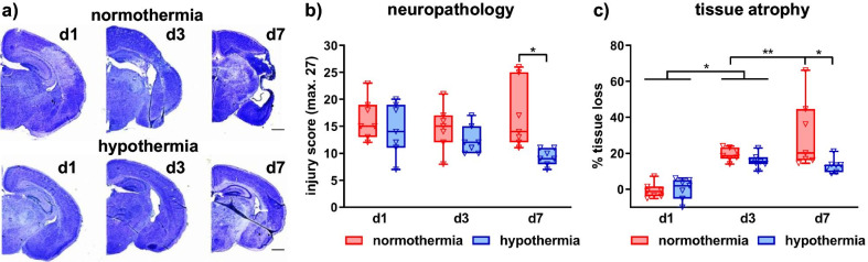
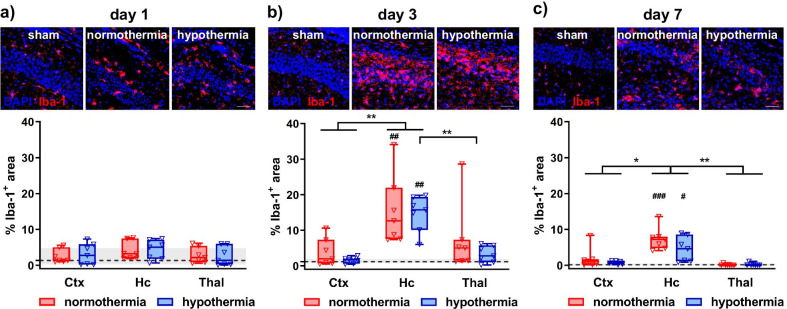
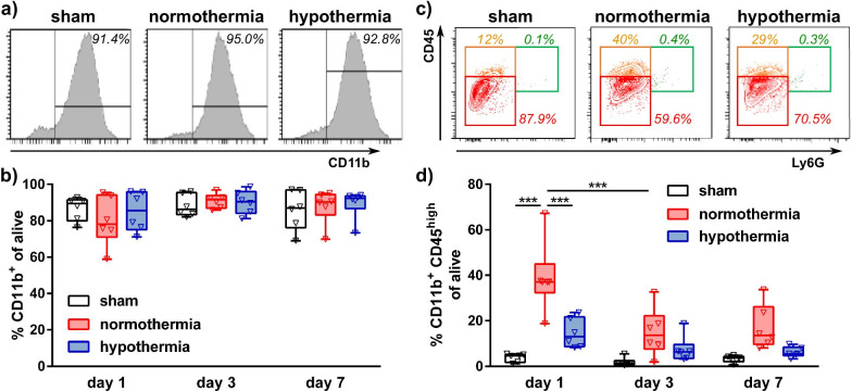
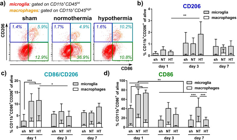
Immediate HT significantly reduced HI-induced brain injury and neuronal loss 7 days post HI, whereas only mild non-significant protection from HI-induced apoptosis and neuronal loss were observed 1 and 3 days after HI. Microglia activation, i.e., Iba-1 immunoreactivity peaked 3 days after HI and was not modulated by HT. However, ex vivo isolated CD11b cells revealed a strong upregulation of the majority of M1 but also M2 marker genes at day 1, which was significantly reduced by HT and rapidly declined at day 3. HI induced a significant increase in the frequency of peripheral macrophages in sorted CD11b cells at day 1, which deteriorated until day 7 and was significantly decreased by HT.
Our data demonstrate that HT-induced neuroprotection is preceded by acute suppression of HI-induced upregulation of inflammatory genes in myeloid cells and decreased infiltration of peripheral macrophages, both representing potential important effector mechanisms of HT.
由于缺氧缺血(HI)导致的新生儿脑病是足月新生儿死亡和残疾的主要原因。治疗性低温(HT)是唯一推荐的治疗方法。然而,仍有 30%的患儿患有神经功能缺陷。炎症是 HI 病理生理学的主要标志,髓样细胞是关键参与者,参与损伤诱导的炎症的进展或消退。在本研究中,我们研究了 HT 对新生 HI 后新生小鼠小胶质细胞/巨噬细胞极化的时空动态的影响。
9 天大的 C57BL/6 小鼠通过阻断右侧颈总动脉后进行 1 小时缺氧来暴露于 HI。HI 后立即进行 4 小时冷却或保持生理体温。在 HI 后 1、3 和 7 天进行分析。通过免疫组织化学评估脑损伤、神经元细胞丢失、细胞凋亡和小胶质细胞激活。通过实时 PCR 分析了广泛的与经典(M1)和替代(M2)髓样细胞激活相关的典型基因,这些基因在离体分离的 CD11b 小胶质细胞/巨噬细胞中进行分析。通过流式细胞术确定分离细胞的纯度和组成。
立即 HT 显著降低了 HI 诱导的脑损伤和神经元丢失,在 HI 后 7 天,而仅观察到对 HI 诱导的细胞凋亡和神经元丢失的轻度非显著性保护,在 HI 后 1 和 3 天。小胶质细胞激活,即 Iba-1 免疫反应性在 HI 后 3 天达到峰值,HT 不调节其表达。然而,离体分离的 CD11b 细胞在第 1 天显示出大多数 M1 但也 M2 标记基因的强烈上调,HT 显著降低了其表达,并且在第 3 天迅速下降。HI 在第 1 天诱导分离的 CD11b 细胞中外周巨噬细胞的频率显著增加,直到第 7 天恶化,并被 HT 显著降低。
我们的数据表明,HT 诱导的神经保护作用之前是髓样细胞中 HI 诱导的炎症基因上调的急性抑制和外周巨噬细胞浸润的减少,这两者都是 HT 的潜在重要效应机制。