Vita-Salute San Raffaele University, Via Olgettina 60, Milan, 20132, Italy.
In Vivo Human Molecular and Structural Neuroimaging Unit, Division of Neuroscience, San Raffaele Scientific Institute, 20132, Milan, Italy.
Alzheimers Res Ther. 2021 Nov 12;13(1):187. doi: 10.1186/s13195-021-00925-1.
Preclinical and pathology evidence suggests an involvement of brain dopamine (DA) circuitry in Alzheimer's disease (AD). We in vivo investigated if, when, and in which target regions [123I]FP-CIT-SPECT regional binding and molecular connectivity are damaged along the AD course.
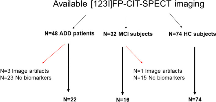
We retrospectively selected 16 amyloid-positive subjects with mild cognitive impairment due to AD (AD-MCI), 22 amyloid-positive patients with probable AD dementia (AD-D), and 74 healthy controls, all with available [123I]FP-CIT-SPECT imaging. We tested whether nigrostriatal vs. mesocorticolimbic dopaminergic targets present binding potential loss, via MANCOVA, and alterations in molecular connectivity, via partial correlation analysis. Results were deemed significant at p < 0.05, after Bonferroni correction for multiple comparisons.
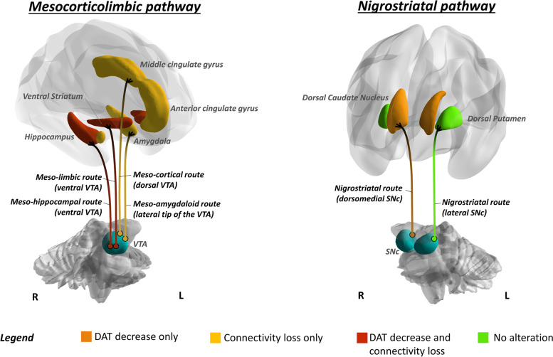
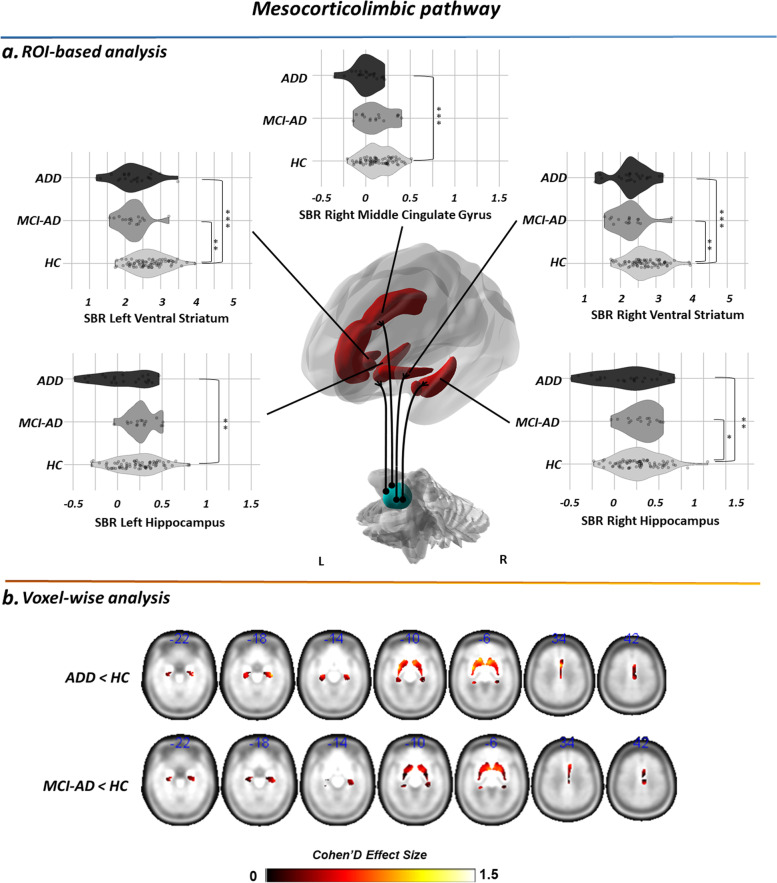
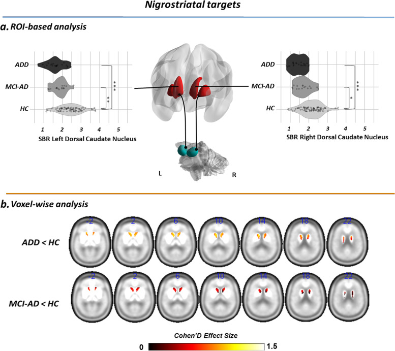
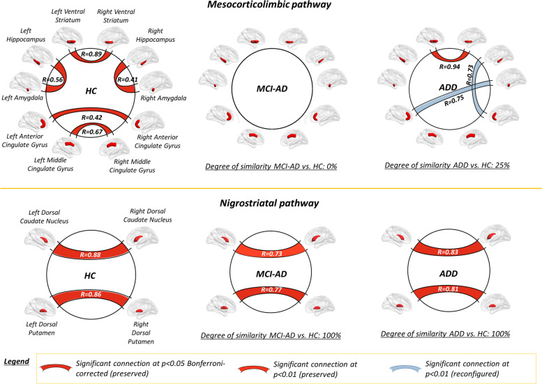
We found significant reductions of [123I]FP-CIT binding in both AD-MCI and AD-D compared to controls. Binding reductions were prominent in the major targets of the ventrotegmental-mesocorticolimbic pathway, namely the ventral striatum and the hippocampus, in both clinical groups, and in the cingulate gyrus, in patients with dementia only. Within the nigrostriatal projections, only the dorsal caudate nucleus showed reduced [123I]FP-CIT binding, in both groups. Molecular connectivity assessment revealed a widespread loss of inter-connections among subcortical and cortical targets of the mesocorticolimbic network only (poor overlap with the control group as expressed by a Dice coefficient ≤ 0.25) and no alterations of the nigrostriatal network (high overlap with controls, Dice coefficient = 1).
Local- and system-level alterations of the mesocorticolimbic dopaminergic circuitry characterize AD, already in prodromal disease phases. These results might foster new therapeutic strategies for AD. The clinical correlates of these findings deserve to be carefully considered within the emergence of both neuropsychiatric symptoms and cognitive deficits.
临床前和病理学证据表明,大脑多巴胺(DA)回路参与了阿尔茨海默病(AD)。我们在体内研究了 AD 进程中[123I]FP-CIT-SPECT 区域结合和分子连接是否以及在哪些靶区受到损伤。
我们回顾性选择了 16 名因 AD 导致轻度认知障碍的淀粉样蛋白阳性患者(AD-MCI)、22 名淀粉样蛋白阳性 AD 痴呆患者(AD-D)和 74 名健康对照者,所有患者均有可用的[123I]FP-CIT-SPECT 成像。我们通过 MANCOVA 测试了纹状体和中皮质边缘多巴胺能靶标是否存在结合潜能损失,通过偏相关分析测试了分子连接的变化。结果在经过 Bonferroni 校正多重比较后,认为 p < 0.05 具有统计学意义。
与对照组相比,我们发现 AD-MCI 和 AD-D 患者的[123I]FP-CIT 结合均显著减少。在两个临床组中,腹侧纹状体和海马等中脑边缘通路的主要靶标均出现结合减少,而在痴呆患者中仅出现扣带回减少。在纹状体投射中,仅背侧尾状核显示[123I]FP-CIT 结合减少,在两个组中均如此。分子连接评估显示,仅中皮质边缘网络的皮质下和皮质靶标之间的连接广泛丧失(与对照组的重叠程度较低,Dice 系数≤0.25),而黑质纹状体网络无改变(与对照组重叠程度较高,Dice 系数=1)。
AD 患者的中皮质边缘多巴胺能回路的局部和系统改变已经在疾病的前驱期表现出来。这些结果可能为 AD 提供新的治疗策略。这些发现的临床相关性值得在神经精神症状和认知缺陷出现时仔细考虑。