Booij J, Tissingh G, Boer G J, Speelman J D, Stoof J C, Janssen A G, Wolters E C, van Royen E A
Graduate School of Neurosciences Amsterdam, The Netherlands.
J Neurol Neurosurg Psychiatry. 1997 Feb;62(2):133-40. doi: 10.1136/jnnp.62.2.133.
The main neuropathological feature in Parkinson's disease is a severe degeneration of the dopaminergic neurons in the substantia nigra resulting in a loss of dopamine (DA) transporters in the striatum. [123I]beta-CIT single photon emission computed tomography (SPECT) studies have demonstrated this loss of striatal DA transporter content in Parkinson's disease in vivo. However, studies with this radioligand also showed that an adequate imaging of the striatal DA transporter content could only be performed on the day after the injection of radioligand, which is not convenient for outpatient evaluations. Recently, a new radioligand [123I]FP-CIT, with faster kinetics than beta-CIT, became available for imaging of the DA transporter with SPECT, and the applicability of this ligand was tested in patients with early and advanced Parkinson's disease, using a one day protocol.
[123I]FP-CIT SPECT was performed in six patients with early and 12 patients with advanced Parkinson's disease, and in six age matched healthy volunteers.
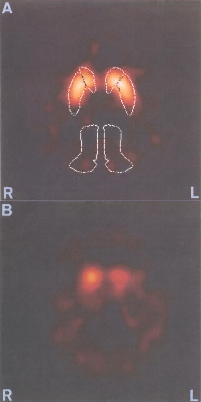
Compared with an age matched control group striatal [123I]FP-CIT uptake in patients with Parkinson's disease was decreased, and this result was measurable three hours after injection of the radioligand. In the Parkinson's disease group the uptake in the putamen was reduced more than in the caudate nucleus. The contralateral striatal uptake of [123I]FP-CIT was significantly lower than the ipsilateral striatal uptake in the Parkinson's disease group. Specific to non-specific striatal uptake ratios correlated with the Hoehn and Yahr stage. A subgroup of patients with early Parkinson's disease also showed significantly lower uptake in the putamen and lower putamen:caudate ratios than controls.
[123I]FP-CIT SPECT allows a significant discrimination between patients with Parkinson's disease and age matched controls with a one day protocol, which will be to great advantage in outpatient evaluations.
帕金森病主要的神经病理学特征是黑质中多巴胺能神经元严重变性,导致纹状体中多巴胺(DA)转运体丧失。[123I]β-CIT单光子发射计算机断层扫描(SPECT)研究已在体内证实帕金森病中纹状体DA转运体含量的这种丧失。然而,使用这种放射性配体的研究还表明,只有在注射放射性配体后的第二天才能对纹状体DA转运体含量进行充分成像,这对于门诊评估来说并不方便。最近,一种新的放射性配体[123I]FP-CIT,其动力学比β-CIT更快,可用于通过SPECT对DA转运体进行成像,并且使用一天方案在早期和晚期帕金森病患者中测试了这种配体的适用性。
对6例早期帕金森病患者、12例晚期帕金森病患者以及6名年龄匹配的健康志愿者进行了[123I]FP-CIT SPECT检查。
与年龄匹配的对照组相比,帕金森病患者纹状体[123I]FP-CIT摄取减少,并且在注射放射性配体3小时后即可检测到这一结果。在帕金森病组中,壳核的摄取减少比尾状核更明显。帕金森病组中,对侧纹状体[123I]FP-CIT摄取明显低于同侧纹状体摄取。特异性与非特异性纹状体摄取比值与Hoehn和Yahr分期相关。一组早期帕金森病患者的壳核摄取也明显低于对照组,并且壳核与尾状核的比值也更低。
[123I]FP-CIT SPECT使用一天方案就能显著区分帕金森病患者和年龄匹配的对照组,这在门诊评估中将具有很大优势。