Department of Radiology, Johns Hopkins University, Baltimore, Maryland, USA.
F.M. Kirby Research Center for Functional Brain Imaging, Kennedy Krieger Institute, Baltimore, Maryland, USA.
NMR Biomed. 2022 Mar;35(3):e4649. doi: 10.1002/nbm.4649. Epub 2021 Nov 15.
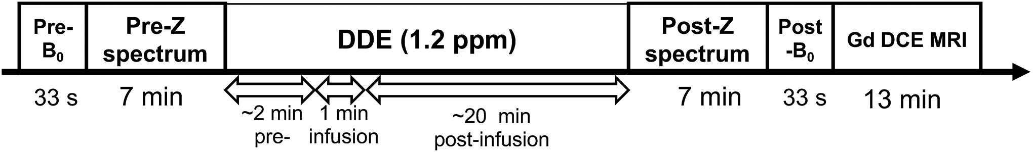
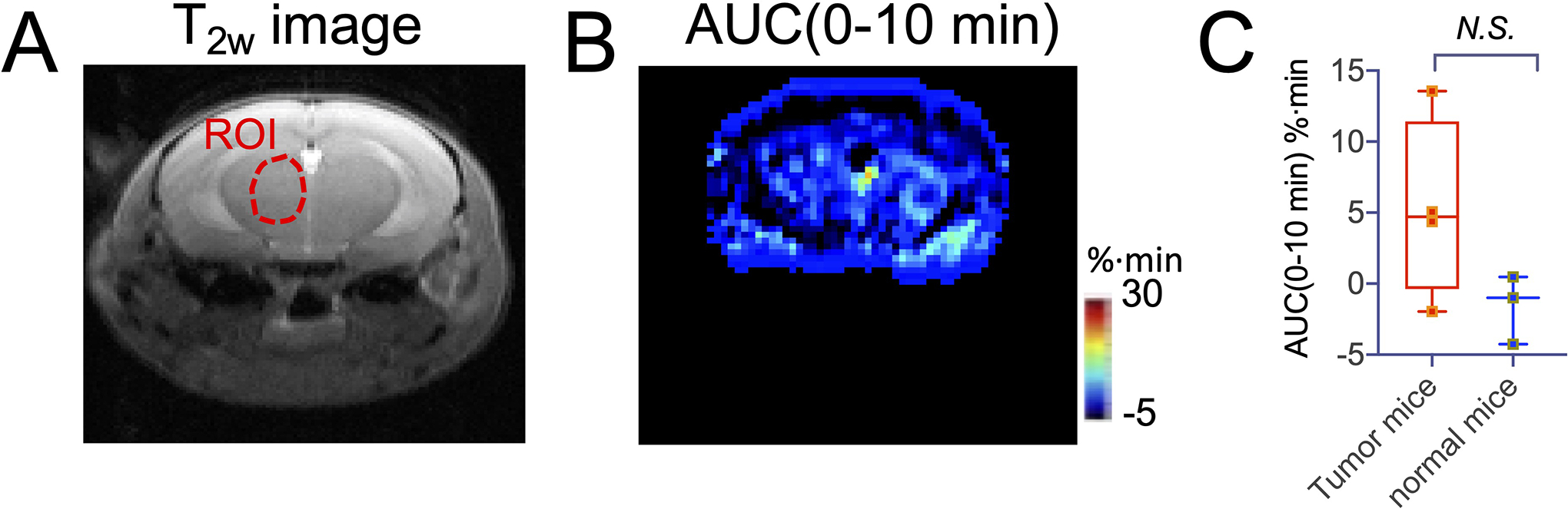
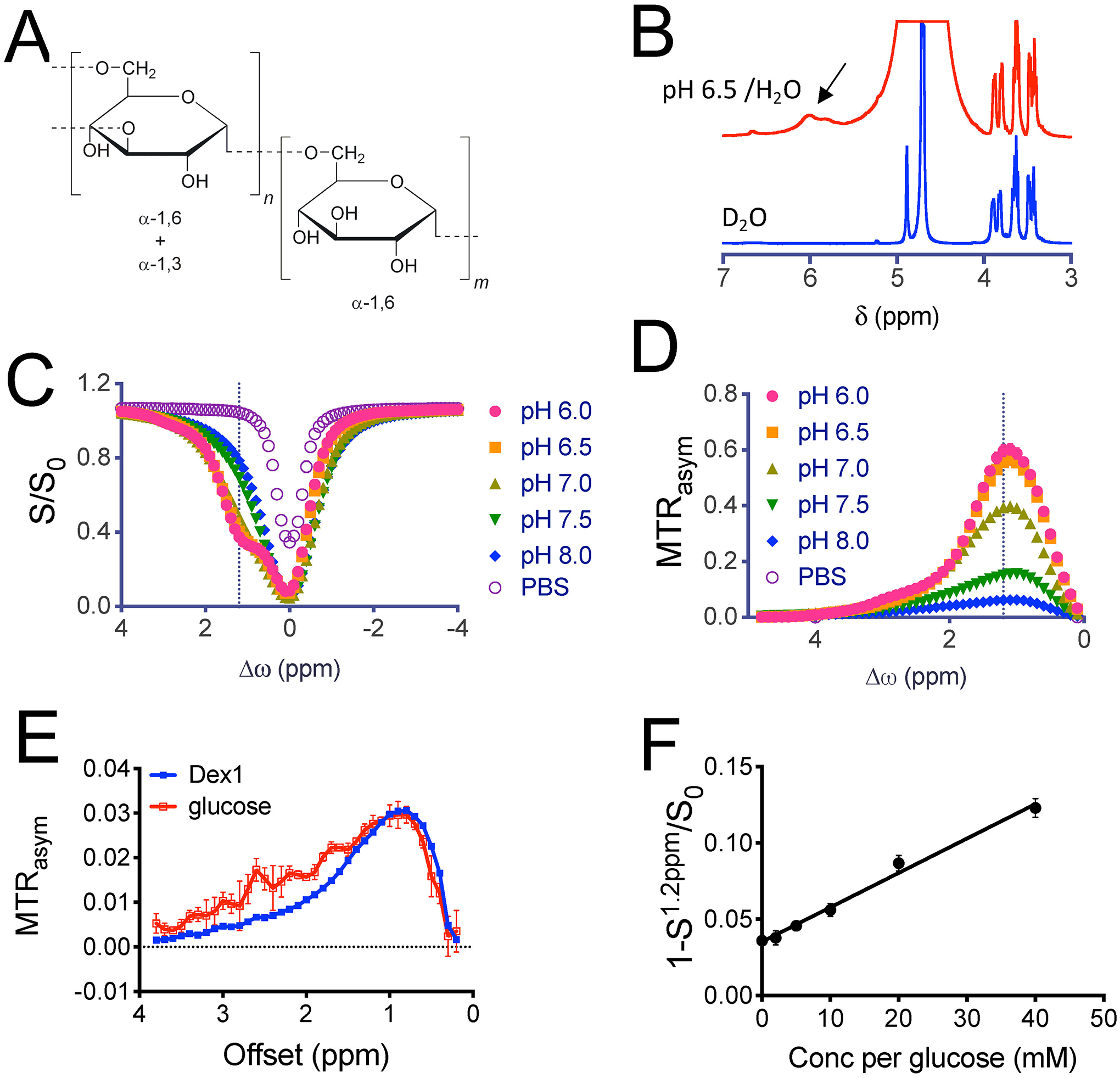
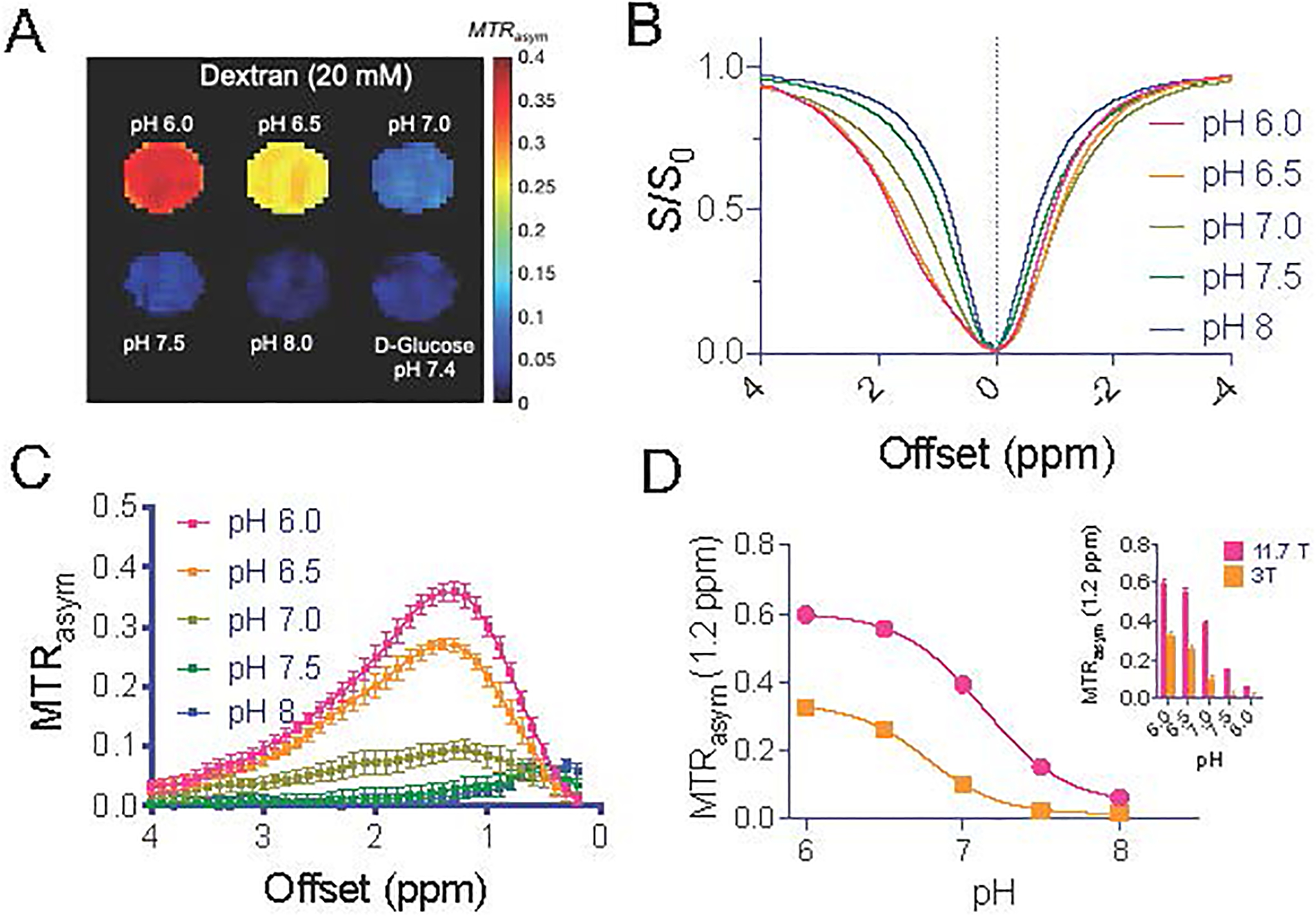
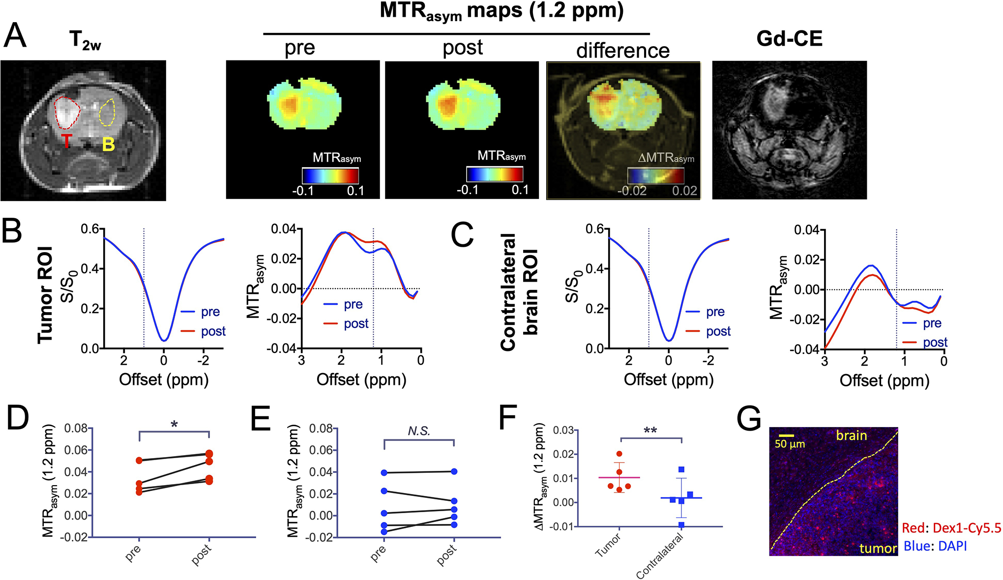
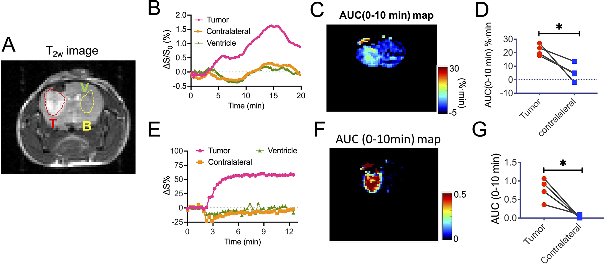
Natural and synthetic sugars have great potential for developing highly biocompatible and translatable chemical exchange saturation transfer (CEST) MRI contrast agents. In this study, we aimed to develop the smallest clinically available form of dextran, Dex1 (molecular weight, MW ~ 1 kDa), as a new CEST agent. We first characterized the CEST properties of Dex1 in vitro at 11.7 T and showed that the Dex1 had a detectable CEST signal at ~1.2 ppm, attributed to hydroxyl protons. In vivo CEST MRI studies were then carried out on C57BL6 mice bearing orthotopic GL261 brain tumors (n = 5) using a Bruker BioSpec 11.7 T MRI scanner. Both steady-state full Z-spectral images and single offset (1.2 ppm) dynamic dextran-enhanced (DDE) images were acquired before and after the intravenous injection of Dex1 (2 g/kg). The steady-state Z-spectral analysis showed a significantly higher CEST contrast enhancement in the tumor than in contralateral brain (∆MTR = 0.010 ± 0.006 versus 0.002 ± 0.008, P = 0.0069) at 20 min after the injection of Dex1. Pharmacokinetic analyses of DDE were performed using the area under the curve (AUC) in the first 10 min after Dex1 injection, revealing a significantly higher uptake of Dex1 in the tumor than in brain tissue for tumor-bearing mice (AUC[0-10 min] = 21.9 ± 4.2 versus 5.3 ± 6.4%·min, P = 0.0294). In contrast, no Dex1 uptake was foundling in the brains of non-tumor-bearing mice (AUC[0-10 min] = -1.59 ± 2.43%·min). Importantly, the CEST MRI findings were consistent with the measurements obtained using DCE MRI and fluorescence microscopy, demonstrating the potential of Dex1 as a highly translatable CEST MRI contrast agent for assessing tumor hemodynamics.
天然和合成糖在开发高度生物相容和可转化的化学交换饱和转移(CEST)MRI 对比剂方面具有巨大潜力。在这项研究中,我们旨在开发最小的临床可用形式的葡聚糖 Dex1(分子量 MW~1 kDa),作为一种新的 CEST 试剂。我们首先在 11.7T 下对 Dex1 的 CEST 特性进行了体外表征,结果表明 Dex1 在约 1.2 ppm 处具有可检测的 CEST 信号,归因于羟基质子。然后,在 11.7T MRI 扫描仪上使用 C57BL6 小鼠进行体内 CEST MRI 研究,该小鼠携带有原位 GL261 脑肿瘤(n=5)。在静脉注射 Dex1(2g/kg)前后,分别采集稳态全 Z 光谱图像和单偏移(1.2 ppm)动态葡聚糖增强(DDE)图像。稳态 Z 光谱分析显示,在注射 Dex1 20 分钟后,肿瘤中的 CEST 对比增强明显高于对侧大脑(∆MTR=0.010±0.006 与 0.002±0.008,P=0.0069)。使用注射 Dex1 后 10 分钟内的曲线下面积(AUC)对 DDE 的药代动力学进行分析,结果显示荷瘤小鼠肿瘤组织对 Dex1 的摄取明显高于脑组织(AUC[0-10 分钟]=21.9±4.2 与 5.3±6.4%·min,P=0.0294)。相比之下,在未患肿瘤的小鼠的大脑中未发现 Dex1 摄取(AUC[0-10 分钟]=-1.59±2.43%·min)。重要的是,CEST MRI 结果与 DCE MRI 和荧光显微镜测量结果一致,表明 Dex1 作为一种高度可转化的 CEST MRI 对比剂,具有评估肿瘤血液动力学的潜力。