Department of Ophthalmology, Kyung Hee University Hospital at Gangdong, 892 Dongnam-ro, Gangdong-gu, Seoul, Republic of Korea.
Barunbit EYE Clinic, Seoul, Republic of Korea.
BMC Ophthalmol. 2021 Nov 18;21(1):399. doi: 10.1186/s12886-021-02152-6.
To identify disease-specific cytokine and growth factor profile differences in the aqueous humor between wet age-related macular degeneration (AMD) patients and age-matched controls and to correlate their levels with the optical coherence tomography (OCT) findings.
Aqueous humors were obtained from 13 wet AMD eyes and 10 control eyes. Twenty cytokines and growth factors were measured using a RayBio antibody microarray technology in wet AMD and control eyes.
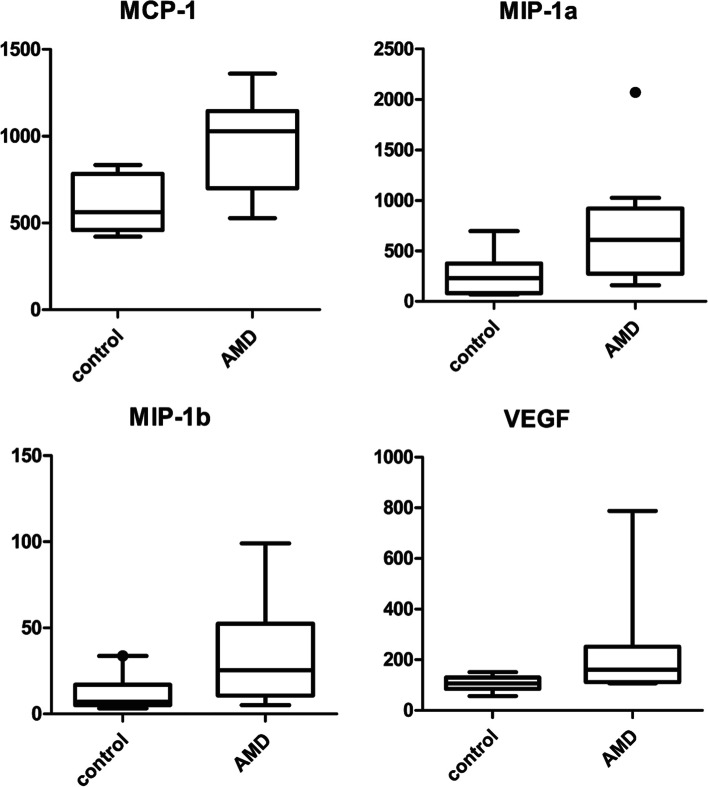
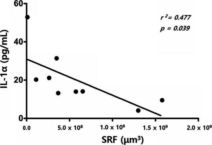
The samples obtained from wet AMD patients exhibited a significantly increased expression of MCP-1, MIP-1α, MIP-1β, and vascular endothelial growth factor (VEGF). Subretinal fluid (SRF) patients showed significantly lower levels of proinflammatory cytokines, such as IL-1α and GM-CSF, than those without SRF. Pigment epithelial detachments (PED) patients showed lower levels of inflammatory cytokines, such as GM-CSF, IFN-γ, and TNF-α, than those without PED. Subretinal tissue (SRT) patients showed a higher level of IFN-γ than those without SRT. Compared with the controls, type 1 macular neovascularization (MNV) patients showed increased levels of MCP-1, MIP-1α, and MIP-1β, but not VEGF (p = 0.083). However, type 2 MNV patients showed increased levels of MCP-1 and VEGF (p = 0.040 and p = 0.040).
Inflammatory cytokines varied according to the type of AMD- and OCT-based parameters. Our observation of low levels of VEGF in patients with type 1 MNV implies that the inhibition of VEGF alone appears to be insufficient treatment for these patients and that cytokines such as MCP-1, MIP-1α, and MIP-1β should be modulated. And the presence of SRF in MNV may be associated with a positive prognosis because we found relatively low levels of proinflammatory cytokines.
为了鉴定湿性年龄相关性黄斑变性(AMD)患者和年龄匹配对照者房水中的特定疾病细胞因子和生长因子谱差异,并将其与光学相干断层扫描(OCT)结果相关联。
从 13 例湿性 AMD 眼和 10 例对照眼获得房水。使用 RayBio 抗体微阵列技术在湿性 AMD 和对照眼中测量了 20 种细胞因子和生长因子。
从湿性 AMD 患者获得的样本显示 MCP-1、MIP-1α、MIP-1β 和血管内皮生长因子(VEGF)的表达显著增加。与无 SRF 的患者相比,有 SRF 的患者的前炎症细胞因子(如 IL-1α 和 GM-CSF)水平显著降低。有 PED 的患者的炎症细胞因子(如 GM-CSF、IFN-γ 和 TNF-α)水平低于无 PED 的患者。与无 SRT 的患者相比,有 SRT 的患者的 IFN-γ 水平较高。与对照组相比,1 型黄斑新生血管(MNV)患者的 MCP-1、MIP-1α 和 MIP-1β 水平升高,但 VEGF 水平未升高(p=0.083)。然而,2 型 MNV 患者的 MCP-1 和 VEGF 水平升高(p=0.040 和 p=0.040)。
根据 AMD 和基于 OCT 的参数的类型,炎症细胞因子会发生变化。我们观察到 1 型 MNV 患者的 VEGF 水平较低,这表明单独抑制 VEGF 似乎不足以治疗这些患者,应该调节 MCP-1、MIP-1α 和 MIP-1β 等细胞因子。并且 MNV 中存在 SRF 可能与良好的预后相关,因为我们发现前炎症细胞因子的水平相对较低。