Townley Ryan A, Botha Hugo, Graff-Radford Jonathan, Whitwell Jennifer, Boeve Bradley F, Machulda Mary M, Fields Julie A, Drubach Daniel A, Savica Rodolfo, Petersen Ronald C, Senjem Matthew L, Knopman David S, Lowe Val J, Jack Clifford R, Josephs Keith A, Jones David T
Department of Neurology, University of Kansas Medical Center, Kansas City, KS 66160, USA.
Department of Neurology, Mayo Clinic, Rochester, MN 55905, USA.
Brain Commun. 2021 Aug 19;3(4):fcab182. doi: 10.1093/braincomms/fcab182. eCollection 2021.
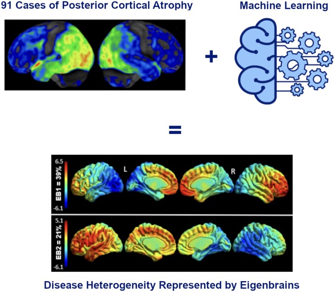
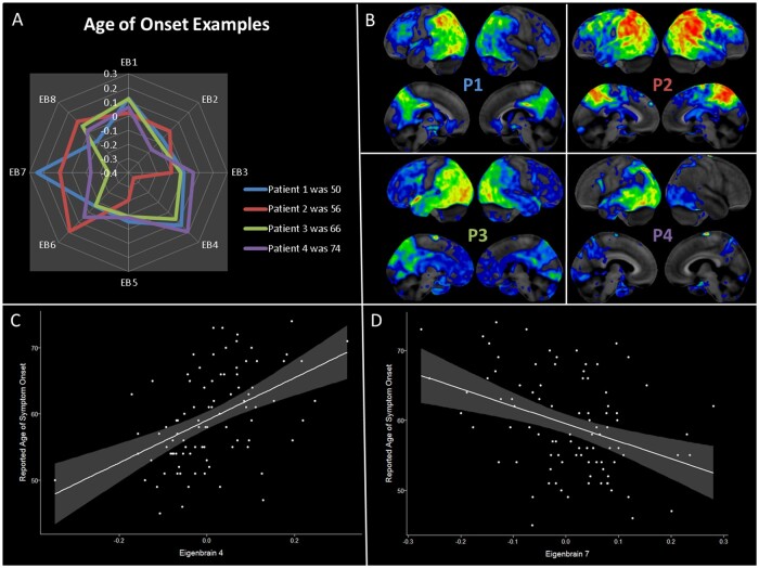
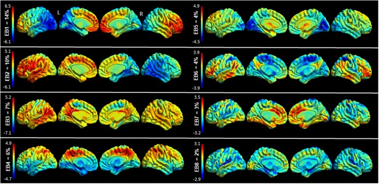
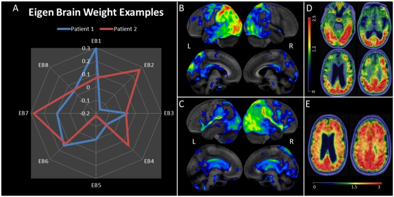
Posterior cortical atrophy is a neurodegenerative syndrome with a heterogeneous clinical presentation due to variable involvement of the left, right, dorsal and ventral parts of the visual system, as well as inconsistent involvement of other cognitive domains and systems. F-fluorodeoxyglucose (FDG)-PET is a sensitive marker for regional brain damage or dysfunction, capable of capturing the pattern of neurodegeneration at the single-participant level. We aimed to leverage these inter-individual differences on FDG-PET imaging to better understand the associations of heterogeneity of posterior cortical atrophy. We identified 91 posterior cortical atrophy participants with FDG-PET data and abstracted demographic, neurologic, neuropsychological and Alzheimer's disease biomarker data. The mean age at reported symptom onset was 59.3 (range: 45-72 years old), with an average disease duration of 4.2 years prior to FDG-PET scan, and a mean education of 15.0 years. Females were more common than males at 1.6:1. After standard preprocessing steps, the FDG-PET scans for the cohort were entered into an unsupervised machine learning algorithm which first creates a high-dimensional space of inter-individual covariance before performing an eigen-decomposition to arrive at a low-dimensional representation. Participant values ('eigenbrains' or latent vectors which represent principle axes of inter-individual variation) were then compared to the clinical and biomarker data. Eight eigenbrains explained over 50% of the inter-individual differences in FDG-PET uptake with left (eigenbrain 1) and right (eigenbrain 2) hemispheric lateralization representing 24% of the variance. Furthermore, eigenbrain-loads mapped onto clinical and neuropsychological data (i.e. aphasia, apraxia and global cognition were associated with the left hemispheric eigenbrain 1 and environmental agnosia and apperceptive prosopagnosia were associated with the right hemispheric eigenbrain 2), suggesting that they captured important axes of normal and abnormal brain function. We used to characterize the eigenbrains through topic-based decoding, which supported the idea that the eigenbrains map onto a diverse set of cognitive functions. These eigenbrains captured important biological and pathophysiologic data (i.e. limbic predominant eigenbrain 4 patterns being associated with older age of onset compared to frontoparietal eigenbrain 7 patterns being associated with younger age of onset), suggesting that approaches that focus on inter-individual differences may be important to better understand the variability observed within a neurodegenerative syndrome like posterior cortical atrophy.
后部皮质萎缩是一种神经退行性综合征,临床表现具有异质性,这是由于视觉系统的左、右、背侧和腹侧部分受累情况不同,以及其他认知领域和系统的受累情况不一致所致。氟脱氧葡萄糖(FDG)-正电子发射断层扫描(PET)是区域脑损伤或功能障碍的敏感标志物,能够在个体水平上捕捉神经退行性变的模式。我们旨在利用FDG-PET成像中的个体差异,更好地理解后部皮质萎缩异质性的关联。我们确定了91名有FDG-PET数据的后部皮质萎缩参与者,并提取了人口统计学、神经学、神经心理学和阿尔茨海默病生物标志物数据。报告症状出现时的平均年龄为59.3岁(范围:45-72岁),在进行FDG-PET扫描前平均病程为4.2年,平均受教育年限为15.0年。女性比男性更常见,比例为1.6:1。经过标准的预处理步骤后,该队列的FDG-PET扫描数据被输入到一种无监督机器学习算法中,该算法首先创建一个个体间协方差的高维空间,然后进行特征分解以获得低维表示。然后将参与者的值(“特征脑”或代表个体间变异主轴线的潜在向量)与临床和生物标志物数据进行比较。八个特征脑解释了FDG-PET摄取中超过50%的个体差异,其中左半球(特征脑1)和右半球(特征脑2)的侧化占变异的24%。此外,特征脑负荷映射到临床和神经心理学数据上(即失语症、失用症和整体认知与左半球特征脑1相关,环境失认症和感知性面孔失认症与右半球特征脑2相关),这表明它们捕捉到了正常和异常脑功能的重要轴线。我们使用基于主题的解码来表征特征脑,这支持了特征脑映射到多种认知功能的观点。这些特征脑捕捉到了重要的生物学和病理生理学数据(即与边缘系统为主的特征脑4模式相比,额顶叶特征脑7模式与发病年龄较小相关),这表明关注个体差异的方法对于更好地理解后部皮质萎缩等神经退行性综合征中观察到的变异性可能很重要。