Pediatric Intensive Care Unit, Children's Hospital of Nanjing Medical University, No 72 Guanzhou Road, Nanjing, China.
Nanjing Infectious Disease Center, The Second Hospital of Nanjing, Nanjing University of Chinese Medicine, Nanjing, China.
World J Pediatr. 2022 Jan;18(1):37-42. doi: 10.1007/s12519-021-00484-3. Epub 2021 Nov 22.
This study aimed to explore the imaging characteristics, diversity and changing trend in CT scans of pediatric patients infected with Delta-variant strain by studying imaging features of children infected with Delta and comparing the results to those of children with original COVID-19.
A retrospective, comparative analysis of initial chest CT manifestations between 63 pediatric patients infected with Delta variant in 2021 and 23 pediatric patients with COVID-19 in 2020 was conducted. Corresponding imaging features were analyzed. In addition, the changing trend in imaging features of COVID-19 Delta-variant cases were explored by evaluating the initial and follow-up CT scans.
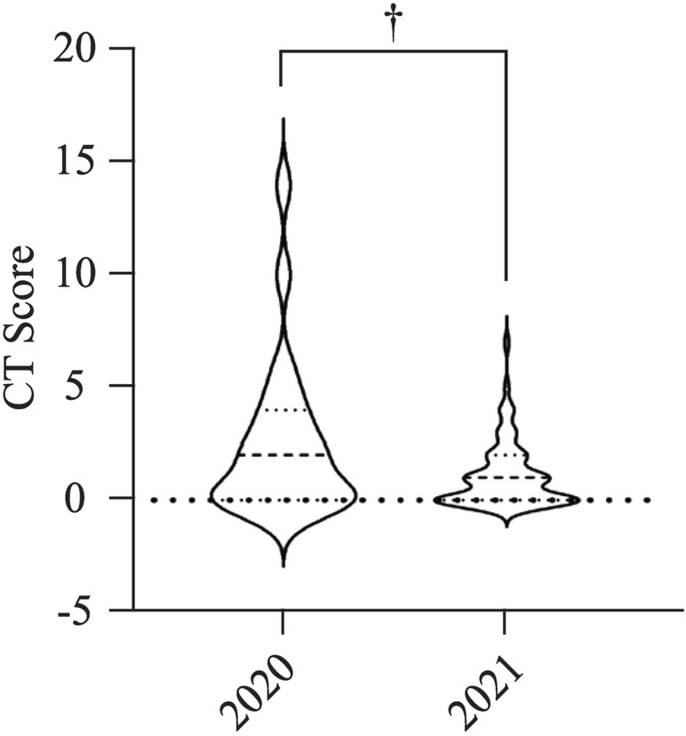
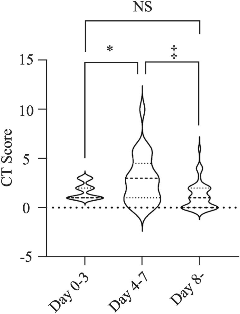
Among 63 children with Delta-variant COVID-19 in 2021, 34 (53.9%) showed positive chest CT presentation; and their CT score (1.10 ± 1.41) was significantly lower than that in 2020 (2.56 ± 3.5) (P = 0.0073). Lesion distribution: lung lesions of Delta cases appear mainly in the lower lungs on both sides. Most children had single lobe involvement (18 cases, 52.9%), 14 (41.2%) in the right lung alone, and 14 (41.2%) in both lungs. A majority of Delta cases displayed initially ground glass (23 cases, 67.6%) and nodular shadows (13 cases, 38.2%) in the first CT scan, with few extrapulmonary manifestations. The 34 children with abnormal chest CT for the first time have a total of 92 chest CT examinations. These children showed a statistically significant difference between the 0-3 day group and the 4-7 day group (P = 0.0392) and a significant difference between the 4-7 day group and the more than 8 days group (P = 0.0003).
The early manifestations of COVID-19 in children with abnormal imaging are mostly small subpleural nodular ground glass opacity. The changes on the Delta-variant COVID-19 chest CT were milder than the original strain. The lesions reached a peak on CT in 4-7 days and quickly improved and absorbed after a week. Dynamic CT re-examination can achieve a good prognosis.
本研究旨在通过研究德尔塔变异株患儿的影像学特征,比较德尔塔变异株患儿与原始 COVID-19 患儿的影像学结果,探讨德尔塔变异株患儿 CT 扫描的影像学特征、多样性和变化趋势。
对 2021 年 63 例德尔塔变异株感染患儿和 2020 年 23 例 COVID-19 患儿的初始胸部 CT 表现进行回顾性、对比分析。分析相应的影像学特征。此外,通过评估初始和随访 CT 扫描,探讨 COVID-19 德尔塔变异株病例影像学特征的变化趋势。
2021 年 63 例德尔塔变异株 COVID-19 患儿中,34 例(53.9%)胸部 CT 表现阳性;其 CT 评分(1.10±1.41)明显低于 2020 年(2.56±3.5)(P=0.0073)。病变分布:德尔塔病例的肺部病变主要出现在双侧下肺。大多数患儿为单叶受累(18 例,52.9%),右肺 14 例(41.2%),双肺 14 例(41.2%)。大多数德尔塔病例在首次 CT 扫描时最初表现为磨玻璃影(23 例,67.6%)和结节影(13 例,38.2%),很少有肺外表现。首次胸部 CT 异常的 34 例患儿共进行了 92 次胸部 CT 检查。0-3 天组和 4-7 天组之间的差异有统计学意义(P=0.0392),4-7 天组和 8 天以上组之间的差异有统计学意义(P=0.0003)。
儿童异常影像学表现的 COVID-19 早期表现多为小胸膜下结节状磨玻璃影。德尔塔变异株 COVID-19 胸部 CT 变化较原始株轻。病变在 4-7 天达到 CT 峰值,一周后迅速改善吸收。动态 CT 复查可取得良好的预后。