Zhong Jiamin, Wang Hao, Yang Ke, Wang Huifeng, Duan Chongwen, Ni Na, An Liqin, Luo Yetao, Zhao Piao, Gou Yannian, Sheng Shiyan, Shi Deyao, Chen Connie, Wagstaff William, Hendren-Santiago Bryce, Haydon Rex C, Luu Hue H, Reid Russell R, Ho Sherwin H, Ameer Guillermo A, Shen Le, He Tong-Chuan, Fan Jiaming
Ministry of Education Key Laboratory of Diagnostic Medicine, And Department of Clinical Biochemistry, School of Laboratory Medicine, Chongqing Medical University, Chongqing, 400016, China.
Molecular Oncology Laboratory, Department of Orthopaedic Surgery and Rehabilitation Medicine, The University of Chicago Medical Center, Chicago, IL, 60637, USA.
Bioact Mater. 2021 Jul 29;9:523-540. doi: 10.1016/j.bioactmat.2021.07.022. eCollection 2022 Mar.
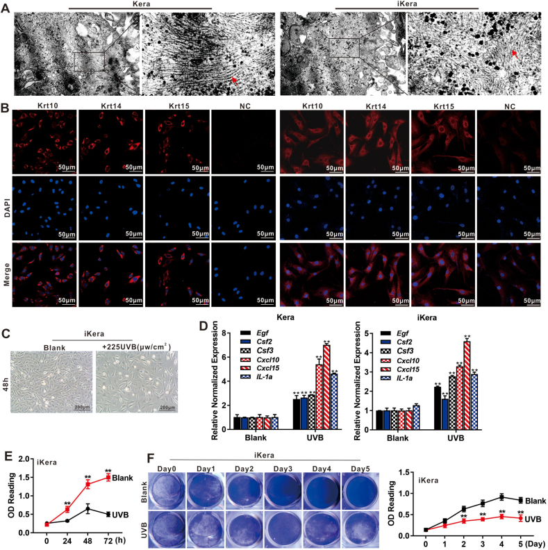
Skin injury is repaired through a multi-phase wound healing process of tissue granulation and re-epithelialization. Any failure in the healing process may lead to chronic non-healing wounds or abnormal scar formation. Although significant progress has been made in developing novel scaffolds and/or cell-based therapeutic strategies to promote wound healing, effective management of large chronic skin wounds remains a clinical challenge. Keratinocytes are critical to re-epithelialization and wound healing. Here, we investigated whether exogenous keratinocytes, in combination with a citrate-based scaffold, enhanced skin wound healing. We first established reversibly immortalized mouse keratinocytes (iKera), and confirmed that the iKera cells expressed keratinocyte markers, and were responsive to UVB treatment, and were non-tumorigenic. In a proof-of-principle experiment, we demonstrated that iKera cells embedded in citrate-based scaffold PPCN provided more effective re-epithelialization and cutaneous wound healing than that of either PPCN or iKera cells alone, in a mouse skin wound model. Thus, these results demonstrate that iKera cells may serve as a valuable skin epithelial source when, combining with appropriate biocompatible scaffolds, to investigate cutaneous wound healing and skin regeneration.
皮肤损伤通过组织肉芽形成和重新上皮化的多阶段伤口愈合过程进行修复。愈合过程中的任何失败都可能导致慢性不愈合伤口或异常瘢痕形成。尽管在开发新型支架和/或基于细胞的治疗策略以促进伤口愈合方面取得了重大进展,但大型慢性皮肤伤口的有效管理仍然是一项临床挑战。角质形成细胞对于重新上皮化和伤口愈合至关重要。在此,我们研究了外源性角质形成细胞与柠檬酸盐基支架联合使用是否能增强皮肤伤口愈合。我们首先建立了可逆永生化小鼠角质形成细胞(iKera),并证实iKera细胞表达角质形成细胞标志物,对UVB治疗有反应,且无致瘤性。在一项原理验证实验中,我们证明在小鼠皮肤伤口模型中,嵌入柠檬酸盐基支架PPCN的iKera细胞比单独的PPCN或iKera细胞能提供更有效的重新上皮化和皮肤伤口愈合。因此,这些结果表明,当与合适的生物相容性支架结合时,iKera细胞可作为一种有价值的皮肤上皮来源,用于研究皮肤伤口愈合和皮肤再生。