Department of Microbiology and Infection Control, Faculty of Medicine, Osaka Medical and Pharmaceutical University, 2-7 Daigaku-machi, Takatsuki, Osaka, 569-8686, Japan.
Med Mol Morphol. 2022 Mar;55(1):60-67. doi: 10.1007/s00795-021-00309-2. Epub 2021 Nov 26.
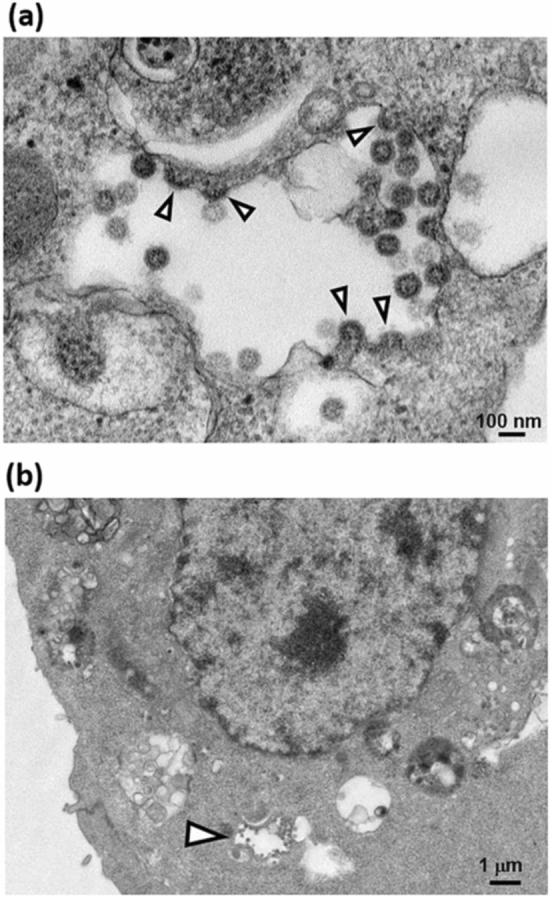
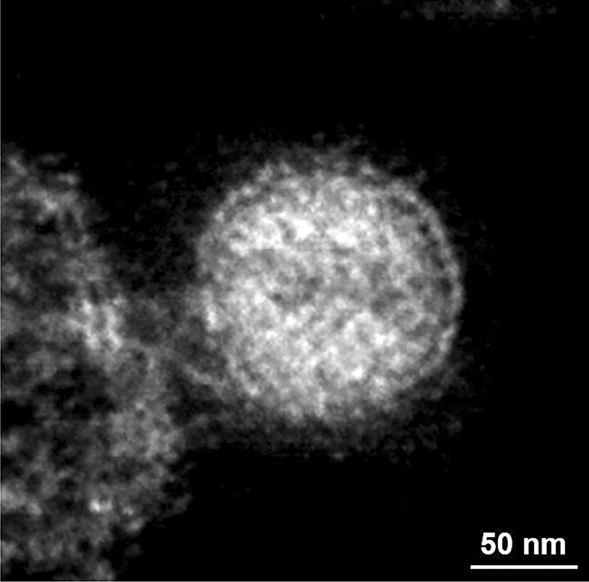
SARS-CoV-2 is the cause of COVID-19. The three-dimensional morphology of viral particles existing and multiplying in infected cells has not been established by electron tomography, which is different from cryo-electron tomography using frozen samples. In this study, we establish the morphological structure of SARS-CoV-2 particles by three-dimensional reconstruction of images obtained by electron tomography and transmission electron microscopy of biological samples embedded in epoxy resin. The characteristic roots of spike structures were found to be arranged at the surface of a virion covered with an envelope. A high-electron-density structure that appears to be a nucleocapsid was observed inside the envelope of the virion on three-dimensional images reconstructed by electron tomography. The SARS-CoV-2 particles that budded in the vacuoles in the cytoplasm were morphologically identical to those found outside the cells, suggesting that mature and infectious SARS-CoV-2 particles were already produced in the vacuoles. Here, we show the three-dimensional morphological structure of SARS-CoV-2 particles reconstructed by electron tomography. To control infection, inhibition of viral release from vacuoles would be a new target in the development of prophylactic agents against SARS-CoV-2.
SARS-CoV-2 是 COVID-19 的病原体。电子断层扫描未能建立存在于感染细胞中并繁殖的病毒粒子的三维形态,这与使用冷冻样本的冷冻电子断层扫描不同。在这项研究中,我们通过对包埋在环氧树脂中的生物样本进行电子断层扫描和透射电子显微镜成像的三维重建,建立了 SARS-CoV-2 粒子的形态结构。发现刺突结构的特征根排列在覆盖包膜的病毒粒子表面。在电子断层扫描重建的三维图像中,观察到包膜内的病毒粒子内部存在一个似乎是核衣壳的高电子密度结构。在细胞质的空泡中出芽的 SARS-CoV-2 粒子在形态上与细胞外的粒子相同,这表明成熟和有感染性的 SARS-CoV-2 粒子已经在空泡中产生。在这里,我们展示了通过电子断层扫描重建的 SARS-CoV-2 粒子的三维形态结构。为了控制感染,抑制病毒从空泡中释放将成为开发针对 SARS-CoV-2 的预防性药物的新靶点。