Shen Chiung-Chyi, Hsu Shan-Hui, Chang Kai-Bo, Yeh Chun-An, Chang Hsiang-Chun, Tang Cheng-Ming, Yang Yi-Chin, Hsieh Hsien-Hsu, Hung Huey-Shan
Department of Neurosurgery, Neurological Institute, Taichung Veterans General Hospital, Taichung 407204, Taiwan.
Department of Physical Therapy, Hung Kuang University, Taichung 433304, Taiwan.
Biomedicines. 2021 Nov 6;9(11):1632. doi: 10.3390/biomedicines9111632.
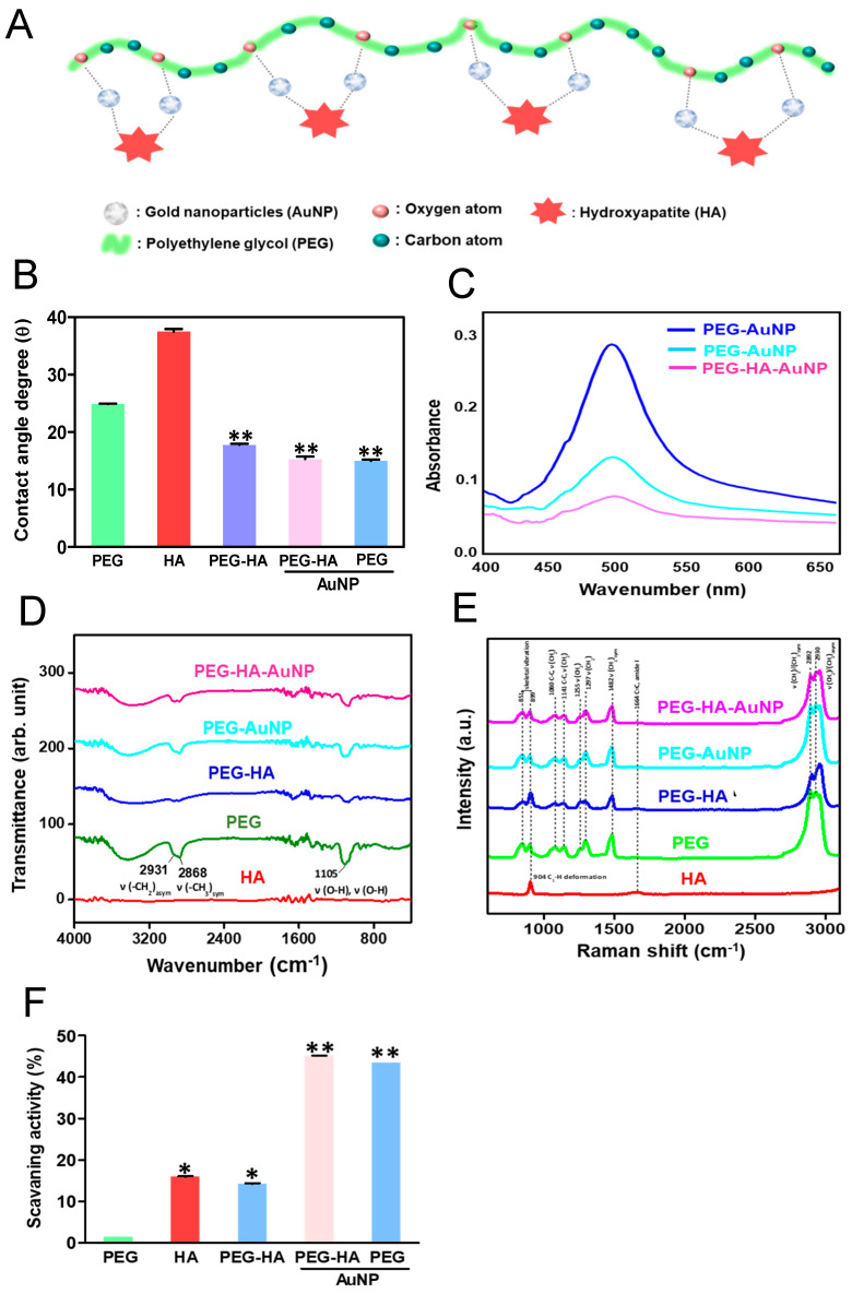
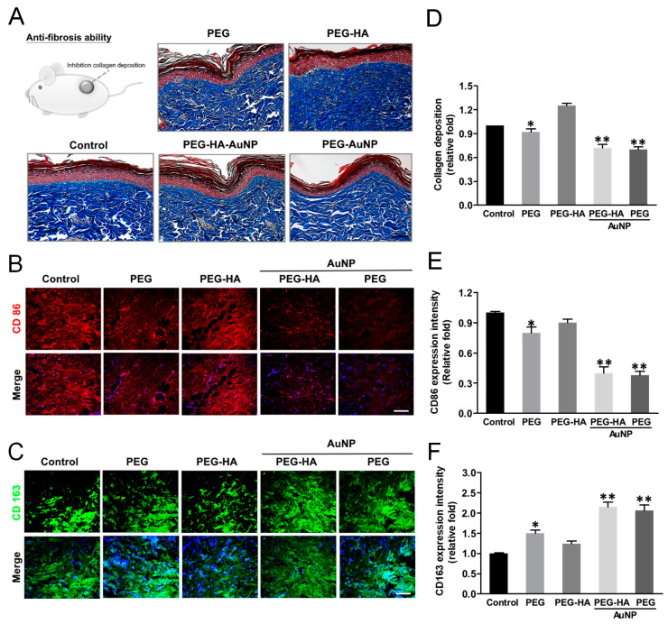
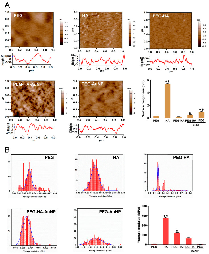
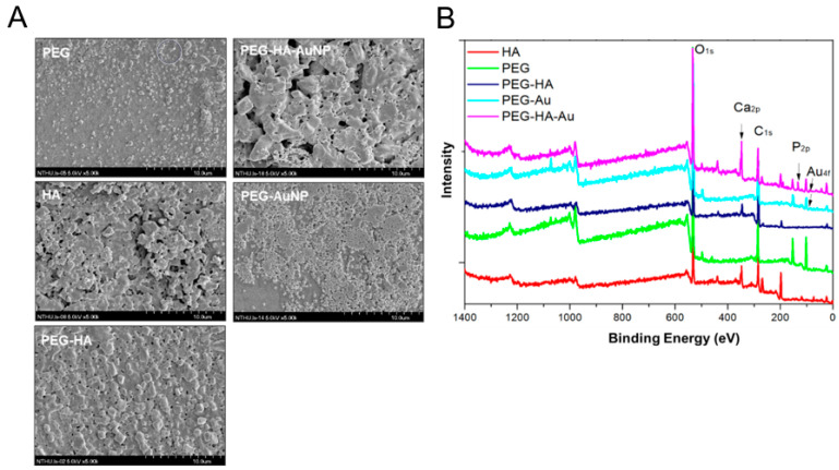
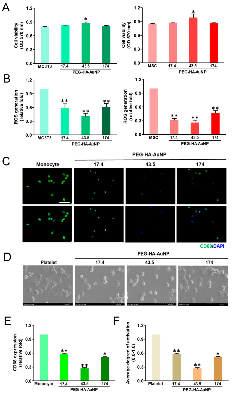
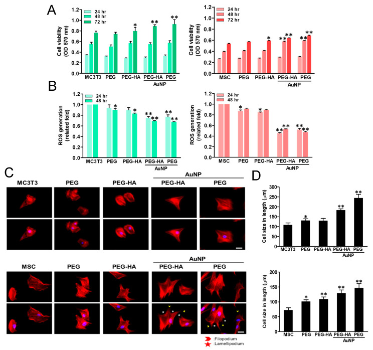
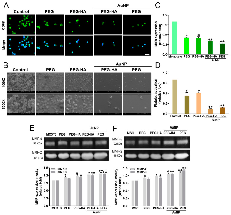
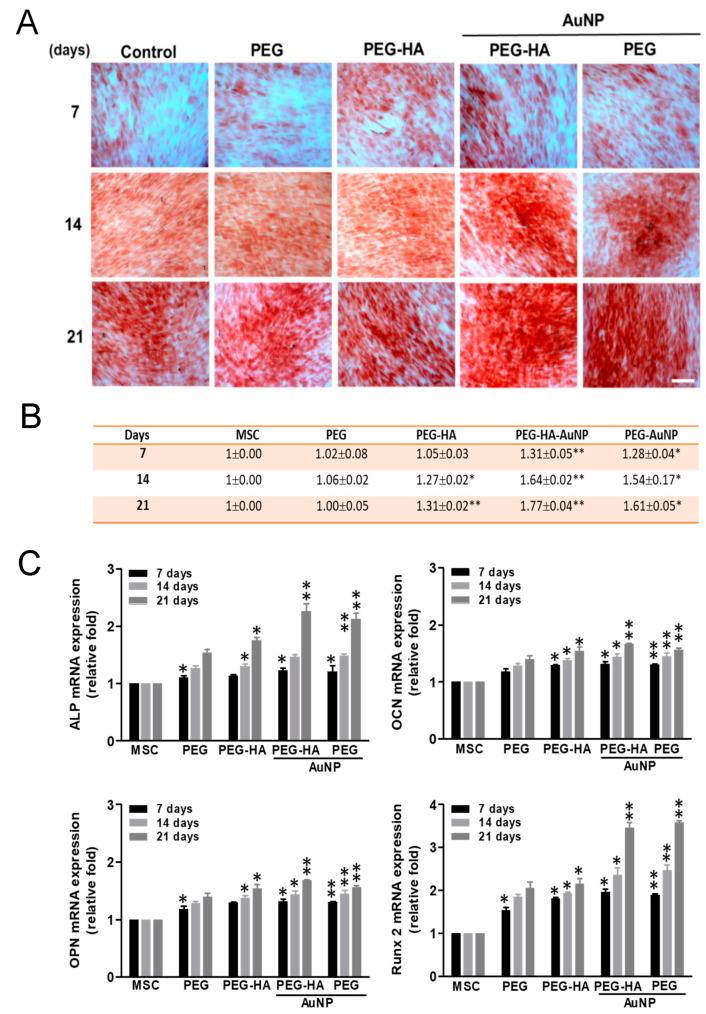
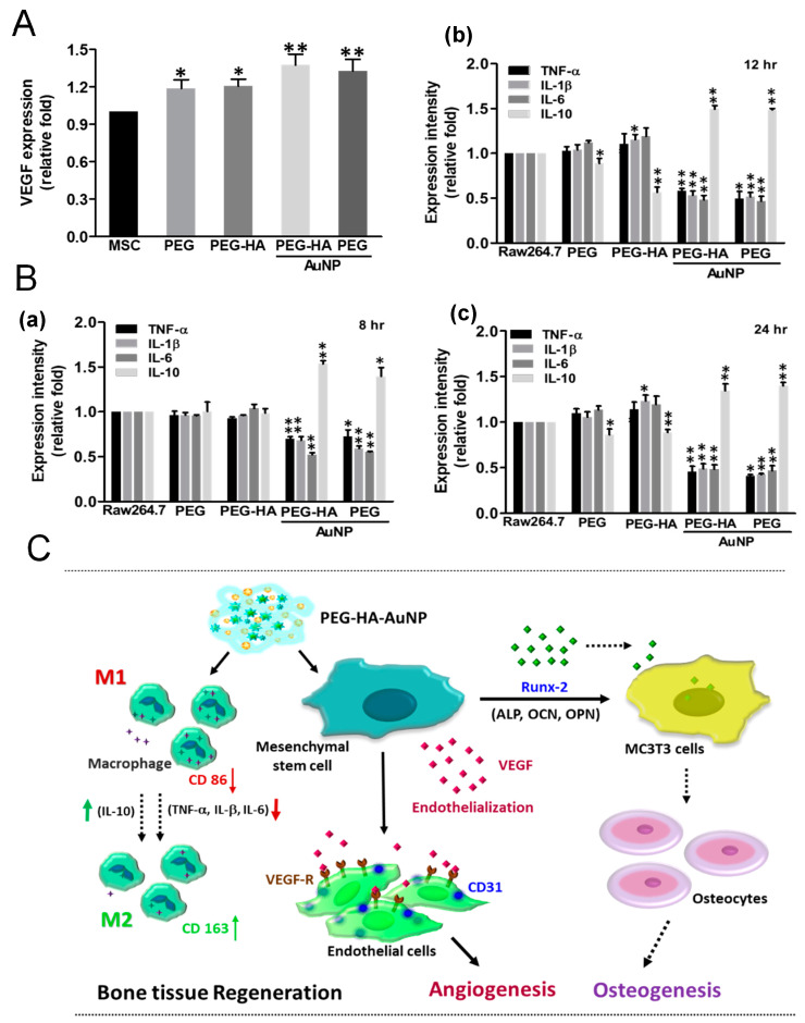
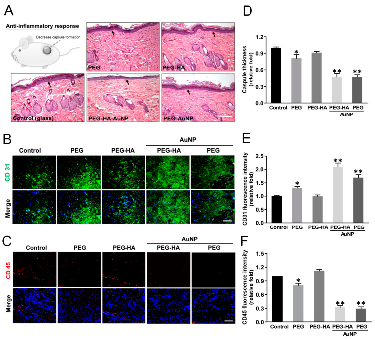
In this study, polyethylene glycol (PEG) with hydroxyapatite (HA), with the incorporation of physical gold nanoparticles (AuNPs), was created and equipped through a surface coating technique in order to form PEG-HA-AuNP nanocomposites. The surface morphology and chemical composition were characterized using scanning electron microscopy (SEM), atomic force microscopy (AFM), UV-Vis spectroscopy (UV-Vis), Fourier transform infrared spectroscopy (FTIR), X-ray photoelectron spectroscopy (XPS), and contact angle assessment. The effects of PEG-HA-AuNP nanocomposites on the biocompatibility and biological activity of MC3T3-E1 osteoblast cells, endothelial cells (EC), macrophages (RAW 264.7), and human mesenchymal stem cells (MSCs), as well as the guiding of osteogenic differentiation, were estimated through the use of an in vitro assay. Moreover, the anti-inflammatory, biocompatibility, and endothelialization capacities were further assessed through in vivo evaluation. The PEG-HA-AuNP nanocomposites showed superior biological properties and biocompatibility capacity for cell behavior in both MC3T3-E1 cells and MSCs. These biological events surrounding the cells could be associated with the activation of adhesion, proliferation, migration, and differentiation processes on the PEG-HA-AuNP nanocomposites. Indeed, the induction of the osteogenic differentiation of MSCs by PEG-HA-AuNP nanocomposites and enhanced mineralization activity were also evidenced in this study. Moreover, from the in vivo assay, we further found that PEG-HA-AuNP nanocomposites not only facilitate the anti-immune response, as well as reducing CD86 expression, but also facilitate the endothelialization ability, as well as promoting CD31 expression, when implanted into rats subcutaneously for a period of 1 month. The current research illustrates the potential of PEG-HA-AuNP nanocomposites when used in combination with MSCs for the regeneration of bone tissue, with their nanotopography being employed as an applicable surface modification approach for the fabrication of biomaterials.
在本研究中,通过表面涂层技术制备并装配了含有羟基磷灰石(HA)的聚乙二醇(PEG),并掺入了物理金纳米颗粒(AuNP),以形成PEG-HA-AuNP纳米复合材料。使用扫描电子显微镜(SEM)、原子力显微镜(AFM)、紫外可见光谱(UV-Vis)、傅里叶变换红外光谱(FTIR)、X射线光电子能谱(XPS)和接触角评估对表面形态和化学成分进行了表征。通过体外试验评估了PEG-HA-AuNP纳米复合材料对MC3T3-E1成骨细胞、内皮细胞(EC)、巨噬细胞(RAW 264.7)和人间充质干细胞(MSC)的生物相容性和生物活性的影响,以及对成骨分化的引导作用。此外,通过体内评估进一步评估了其抗炎、生物相容性和内皮化能力。PEG-HA-AuNP纳米复合材料在MC3T3-E1细胞和MSC中均表现出优异的生物学特性和对细胞行为的生物相容性能力。围绕细胞的这些生物学事件可能与PEG-HA-AuNP纳米复合材料上粘附、增殖、迁移和分化过程的激活有关。事实上,本研究还证实了PEG-HA-AuNP纳米复合材料对MSC成骨分化的诱导作用和增强的矿化活性。此外,从体内试验中,我们进一步发现,当将PEG-HA-AuNP纳米复合材料皮下植入大鼠体内1个月时,它不仅促进免疫反应,降低CD86表达,还促进内皮化能力,提高CD31表达。当前的研究说明了PEG-HA-AuNP纳米复合材料与MSC联合用于骨组织再生的潜力,其纳米拓扑结构可作为一种适用的表面改性方法用于生物材料的制造。