Department of Ophthalmology, CHA Bundang Medical Center, CHA University, Seongnam 13496, Gyeonggi-do, Korea.
Department of Biomedical Science, CHA University, Seongnam 13488, Gyeonggi-do, Korea.
Int J Mol Sci. 2021 Nov 20;22(22):12529. doi: 10.3390/ijms222212529.
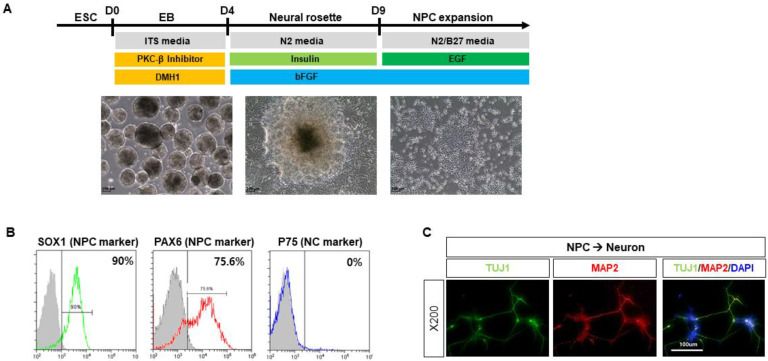
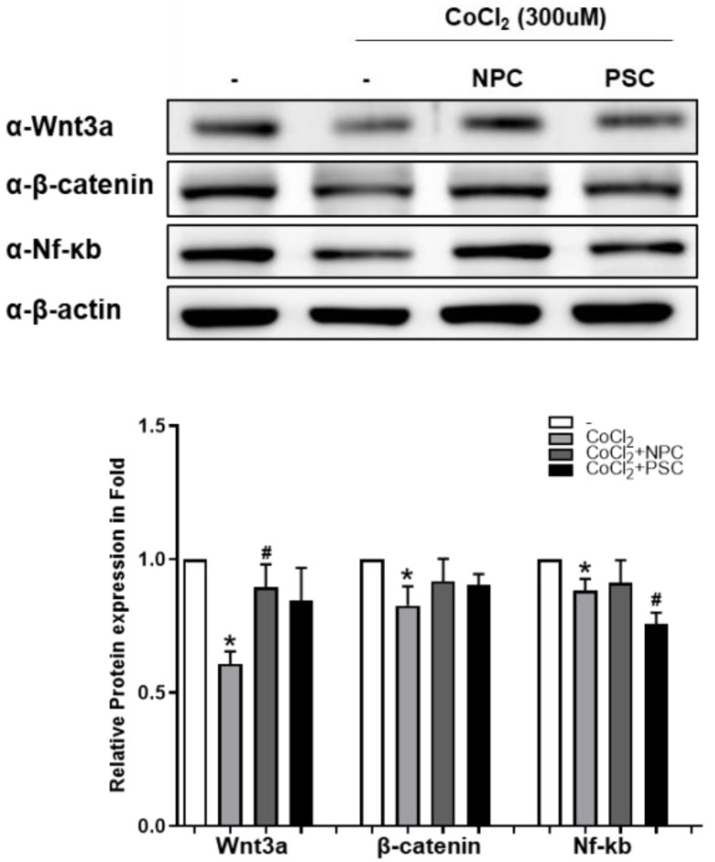
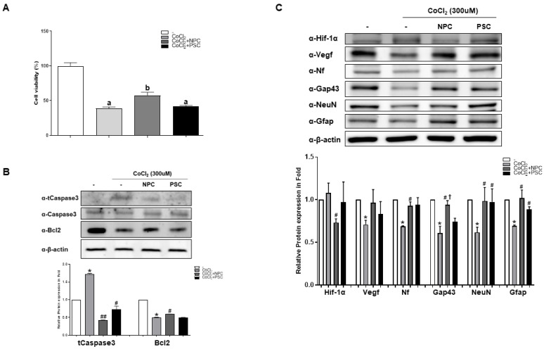
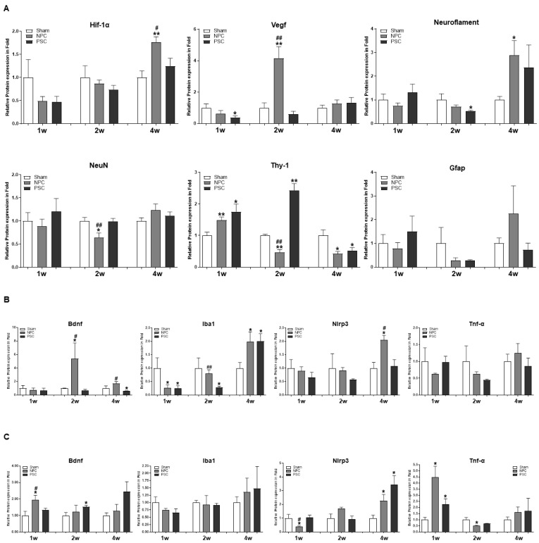
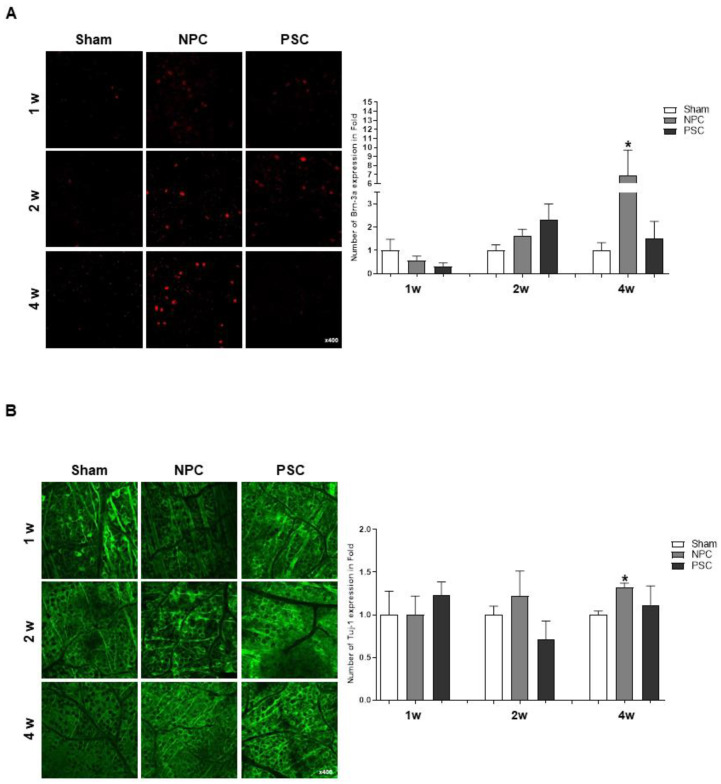
Human pluripotent stem cell-derived neural progenitor cells (NPCs) have the potential to recover from nerve injury. We previously reported that human placenta-derived mesenchymal stem cells (PSCs) have neuroprotective effects. To evaluate the potential benefit of NPCs, we compared them to PSCs using R28 cells under hypoxic conditions and a rat model of optic nerve injury. NPCs and PSCs (2 × 10⁶ cells) were injected into the subtenon space. After 1, 2, and 4 weeks, we examined changes in target proteins in the retina and optic nerve. NPCs significantly induced vascular endothelial growth factor (Vegf) compared to age-matched shams and PSC groups at 2 weeks; they also induced neurofilaments in the retina compared to the sham group at 4 weeks. In addition, the expression of brain-derived neurotrophic factor (Bdnf) was high in the retina in the NPC group at 2 weeks, while expression in the optic nerve was high in both the NPC and PSC groups. The low expression of ionized calcium-binding adapter molecule 1 (Iba1) in the retina had recovered at 2 weeks after NPC injection and at 4 weeks after PSC injection. The expression of the inflammatory protein NLR family, pyrin domain containing 3 (Nlrp3) was significantly reduced at 1 week, and that of tumor necrosis factor-α (Tnf-α) in the optic nerves of the NPC group was lower at 2 weeks. Regarding retinal ganglion cells, the expressions of Brn3a and Tuj1 in the retina were enhanced in the NPC group compared to sham controls at 4 weeks. NPC injections increased Gap43 expression from 2 weeks and reduced Iba1 expression in the optic nerves during the recovery period. In addition, R28 cells exposed to hypoxic conditions showed increased cell survival when cocultured with NPCs compared to PSCs. Both Wnt/β-catenin signaling and increased Nf-ĸb could contribute to the rescue of damaged retinal ganglion cells via upregulation of neuroprotective factors, microglial engagement, and anti-inflammatory regulation by NPCs. This study suggests that NPCs could be useful for the cellular treatment of various optic neuropathies, together with cell therapy using mesenchymal stem cells.
人多能干细胞衍生的神经祖细胞(NPCs)具有从神经损伤中恢复的潜力。我们之前报道过,人胎盘来源的间充质干细胞(PSCs)具有神经保护作用。为了评估 NPCs 的潜在益处,我们在缺氧条件下和大鼠视神经损伤模型中,将 NPCs 与 PSCs 进行了比较。将 NPCs 和 PSCs(2×10⁶ 个细胞)注射到腱膜下间隙。在 1、2 和 4 周后,我们检查了视网膜和视神经中靶蛋白的变化。与年龄匹配的假手术组和 PSC 组相比,NPCs 在 2 周时显著诱导血管内皮生长因子(Vegf);与假手术组相比,NPCs 在 4 周时还诱导了视网膜中的神经丝。此外,在 2 周时,NPC 组的视网膜中脑源性神经营养因子(Bdnf)表达较高,而在 NPC 和 PSC 组的视神经中表达较高。NPC 注射后 2 周和 PSC 注射后 4 周,视网膜中离子钙结合接头分子 1(Iba1)的低表达得到恢复。在 1 周时,炎症蛋白 NLR 家族、含 pyrin 域的蛋白 3(Nlrp3)的表达显著降低,在 2 周时,NPC 组视神经中的肿瘤坏死因子-α(Tnf-α)表达降低。关于视网膜神经节细胞,与假手术对照组相比,NPC 组在 4 周时视网膜中的 Brn3a 和 Tuj1 表达增强。NPC 注射后,从第 2 周开始,Gap43 表达增加,在恢复期间,视神经中的 Iba1 表达减少。此外,与 PSCs 共培养时,暴露于缺氧条件下的 R28 细胞的细胞存活率增加。Wnt/β-catenin 信号通路和 NF-ĸB 的增加可能通过上调神经营养因子、小胶质细胞的参与以及 NPC 对炎症的调节来促进损伤的视网膜神经节细胞的恢复。这项研究表明,NPCs 可与间充质干细胞的细胞治疗一起,用于各种视神经病变的细胞治疗。