Rozza Ariane Leite, Beserra Fernando Pereira, Vieira Ana Júlia, Oliveira de Souza Eduardo, Hussni Carlos Alberto, Martinez Emanuel Ricardo Monteiro, Nóbrega Rafael Henrique, Pellizzon Cláudia Helena
Department of Structural and Functional Biology, Institute of Biosciences, São Paulo State University (UNESP), Dr. Antonio Celso W Zanin Street, 250, Botucatu 18618-689, Brazil.
Department of Surgery and Veterinary Anesthesiology, School of Veterinary Medicine and Animal Science, São Paulo State University (UNESP), Dr. Walter M Correa Street, Botucatu 18618-689, Brazil.
Pharmaceutics. 2021 Nov 9;13(11):1902. doi: 10.3390/pharmaceutics13111902.
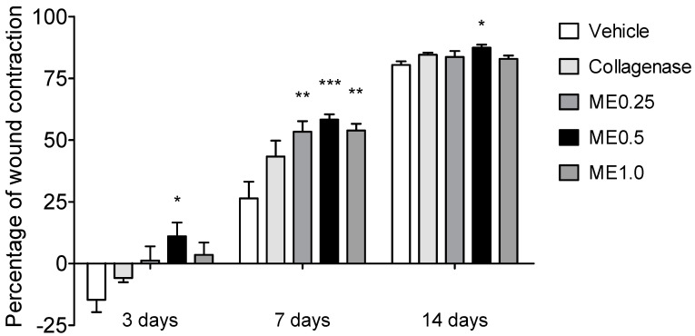
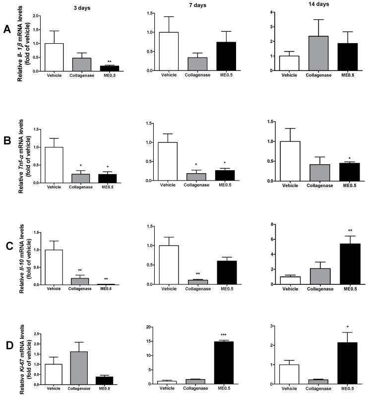
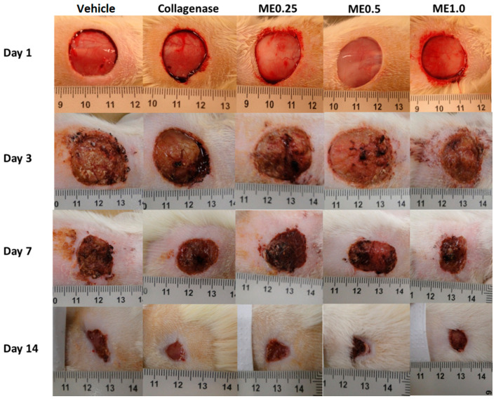
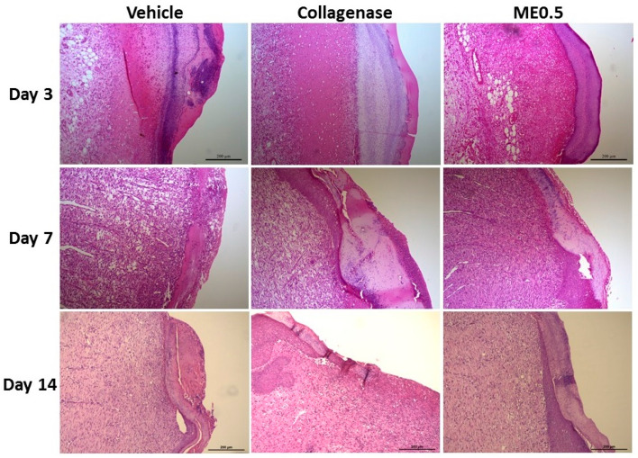
Wound healing involves inflammatory, proliferative, and remodeling phases, in which various cells and chemical intermediates are involved. This study aimed to investigate the skin wound healing potential of menthol, as well as the mechanisms involved in its effect, after 3, 7, or 14 days of treatment, according to the phases of wound healing. Skin wound was performed in the back of Wistar rats, which were topically treated with vehicle cream; collagenase-based cream (1.2 U/g); or menthol-based cream at 0.25%, 0.5%, or 1.0% over 3, 7, or 14 days. Menthol cream at 0.5% accelerated the healing right from the inflammatory phase (3 days) by decreasing mRNA expression of inflammatory cytokines TNF-α and Il-6. At the proliferative phase (7 days), menthol 0.5% increased the activity of antioxidant enzymes SOD, GR, and GPx, as well as the level of GSH, in addition to decreasing the levels of inflammatory cytokines TNF-α, IL-6, and IL-1β and augmenting mRNA expression for Ki-67, a marker of cellular proliferation. At the remodeling phase (14 days), levels of inflammatory cytokines were decreased, and the level of Il-10 and its mRNA expression were increased in the menthol 0.5% group. Menthol presented skin wound healing activity by modulating the antioxidant system of the cells and the inflammatory response, in addition to stimulating epithelialization.
伤口愈合涉及炎症、增殖和重塑阶段,其中涉及多种细胞和化学中间体。本研究旨在根据伤口愈合阶段,研究薄荷醇在治疗3、7或14天后的皮肤伤口愈合潜力及其作用机制。在Wistar大鼠背部制造皮肤伤口,在3、7或14天内分别用赋形剂乳膏、含1.2 U/g胶原酶的乳膏或含0.25%、0.5%或1.0%薄荷醇的乳膏进行局部治疗。0.5%的薄荷醇乳膏从炎症期(3天)开始就通过降低炎症细胞因子TNF-α和Il-6的mRNA表达来加速愈合。在增殖期(7天),0.5%的薄荷醇除了降低炎症细胞因子TNF-α、IL-6和IL-1β的水平并增加细胞增殖标志物Ki-67的mRNA表达外,还提高了抗氧化酶SOD、GR和GPx的活性以及GSH水平。在重塑期(14天),0.5%薄荷醇组的炎症细胞因子水平降低,Il-10水平及其mRNA表达增加。薄荷醇除了刺激上皮形成外,还通过调节细胞的抗氧化系统和炎症反应来呈现皮肤伤口愈合活性。