• 文献检索
  • 文档翻译
  • 深度研究
  • 学术资讯
  • Suppr Zotero 插件Zotero 插件
  • 邀请有礼
  • 套餐&价格
  • 历史记录
应用&插件
Suppr Zotero 插件Zotero 插件浏览器插件Mac 客户端Windows 客户端微信小程序
定价
高级版会员购买积分包购买API积分包
服务
文献检索文档翻译深度研究API 文档MCP 服务
关于我们
关于 Suppr公司介绍联系我们用户协议隐私条款
关注我们

Suppr 超能文献

核心技术专利:CN118964589B侵权必究
粤ICP备2023148730 号-1Suppr @ 2026

文献检索

告别复杂PubMed语法,用中文像聊天一样搜索,搜遍4000万医学文献。AI智能推荐,让科研检索更轻松。

立即免费搜索

文件翻译

保留排版,准确专业,支持PDF/Word/PPT等文件格式,支持 12+语言互译。

免费翻译文档

深度研究

AI帮你快速写综述,25分钟生成高质量综述,智能提取关键信息,辅助科研写作。

立即免费体验

前房 PRESERFLO™ 微分流植入术后角膜内皮细胞丢失:前节光学相干断层扫描管位置作为一个危险因素

Corneal Endothelial Cell Loss After PRESERFLO™ MicroShunt Implantation in the Anterior Chamber: Anterior Segment OCT Tube Location as a Risk Factor.

作者信息

Ibarz-Barberá Marta, Morales-Fernández Laura, Corroto-Cuadrado Arturo, Martinez-Galdón Fátima, Tañá-Rivero Pedro, Gómez de Liaño Rosario, Teus Miguel A

机构信息

Grupo Oftalvist, Juan Bravo Street #1, 28006, Madrid, Spain.

Hospital Moncloa, HLA Hospitales, Madrid, Spain.

出版信息

Ophthalmol Ther. 2022 Feb;11(1):293-310. doi: 10.1007/s40123-021-00428-0. Epub 2021 Nov 26.

DOI:10.1007/s40123-021-00428-0
PMID:34837167
原文链接:https://pmc.ncbi.nlm.nih.gov/articles/PMC8770787/
Abstract

INTRODUCTION

To analyze the effects of PRESERFLO on corneal endothelial cell density (ECD).

METHODS

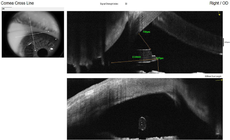
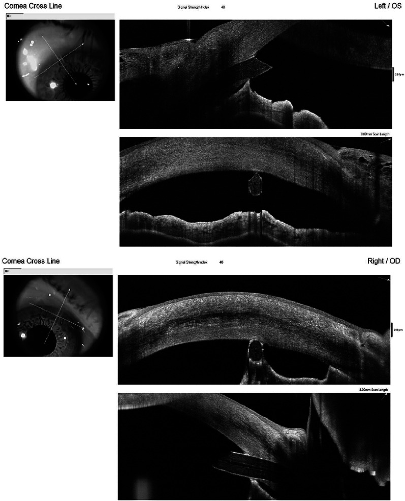
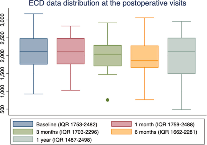
Forty-six eyes that underwent PRESERFLO implantation were followed up for 12 months. Specular microscopy was performed preoperatively and at 1, 3, 6, and 12 months postoperatively to measure central ECD and mean monthly reduction (MMR). Anterior segment optical coherence tomography (AS-OCT) was applied to measure the tube-endothelium (TE < 200 μm, 201-500 μm, > 500 μm) distance. The relationship between TE distance and ECD was analyzed with a linear mixed-effects model.

RESULTS

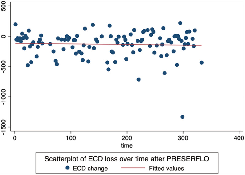
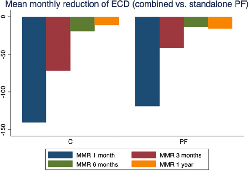
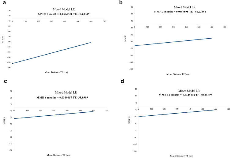
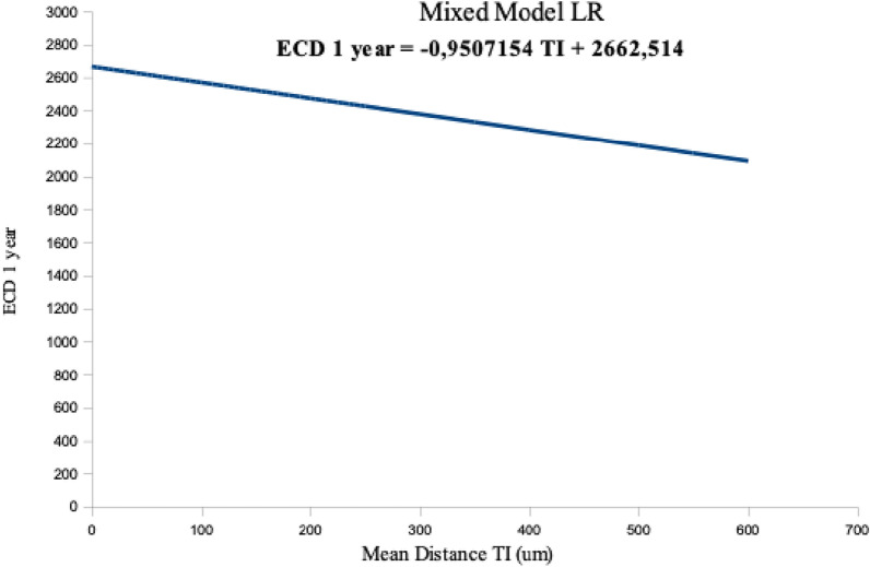
Central ECD decreased significantly at 1 year (7.4%, p = 0.04), with an MMR of -15 ± 25 cells/mm. Regarding TE distance groups, there was an 18% ECD reduction in the < 200 μm group vs. 1% in the > 500 μm group (p = 0.08). Endothelial cell loss was related to TE distance (mean 482.9 ± 238 μm), with a higher rate at 1 month in comparison to 12 months for the same tube position in the anterior chamber (-174.8 ± 65.2 cells/mm at 1 month vs. 30.2 ± 11.3 cells/mm at 12 months, p < 0.01). From month 6, tubes located > 600 μm from the endothelium showed EC loss close to zero.

CONCLUSIONS

The PRESERFLO implant is associated with a loss of EC from the immediate postoperative period that continues over time at lower rates. A shorter TE distance appears to cause more severe ECD loss.

摘要

引言

分析 PRESERFLO 对角膜内皮细胞密度(ECD)的影响。

方法

对 46 只接受 PRESERFLO 植入术的眼睛进行了 12 个月的随访。术前以及术后 1、3、6 和 12 个月进行镜面显微镜检查,以测量中央 ECD 和平均每月降低值(MMR)。应用眼前节光学相干断层扫描(AS-OCT)测量管-内皮(TE<200μm、201 - 500μm、>500μm)距离。使用线性混合效应模型分析 TE 距离与 ECD 之间的关系。

结果

1 年时中央 ECD 显著下降(7.4%,p = 0.04),MMR 为 -15±25 个细胞/mm²。关于 TE 距离组,<200μm 组的 ECD 降低了 18%,而>500μm 组为 1%(p = 0.08)。内皮细胞丢失与 TE 距离有关(平均 482.9±238μm),在前房相同管位置,1 个月时的丢失率高于 12 个月时(1 个月时为 -174.8±65.2 个细胞/mm²,12 个月时为 30.2±11.3 个细胞/mm²,p<0.01)。从第 6 个月起,距离内皮>600μm 的管显示内皮细胞丢失接近零。

结论

PRESERFLO 植入物与术后即刻开始的内皮细胞丢失有关,且随着时间推移以较低速率持续。较短的 TE 距离似乎会导致更严重的 ECD 丢失。

https://cdn.ncbi.nlm.nih.gov/pmc/blobs/2baf/8770787/9d142db2409d/40123_2021_428_Fig10_HTML.jpg
https://cdn.ncbi.nlm.nih.gov/pmc/blobs/2baf/8770787/8e12c6c94f8f/40123_2021_428_Fig1_HTML.jpg
https://cdn.ncbi.nlm.nih.gov/pmc/blobs/2baf/8770787/09bb2d63d8b8/40123_2021_428_Fig2_HTML.jpg
https://cdn.ncbi.nlm.nih.gov/pmc/blobs/2baf/8770787/a74cf77f45d6/40123_2021_428_Fig3_HTML.jpg
https://cdn.ncbi.nlm.nih.gov/pmc/blobs/2baf/8770787/9bbe1d52236d/40123_2021_428_Fig4_HTML.jpg
https://cdn.ncbi.nlm.nih.gov/pmc/blobs/2baf/8770787/e74a582a739b/40123_2021_428_Fig5_HTML.jpg
https://cdn.ncbi.nlm.nih.gov/pmc/blobs/2baf/8770787/05b10da249f7/40123_2021_428_Fig6_HTML.jpg
https://cdn.ncbi.nlm.nih.gov/pmc/blobs/2baf/8770787/b05fa4f7b605/40123_2021_428_Fig7_HTML.jpg
https://cdn.ncbi.nlm.nih.gov/pmc/blobs/2baf/8770787/3e01337c45d2/40123_2021_428_Fig8_HTML.jpg
https://cdn.ncbi.nlm.nih.gov/pmc/blobs/2baf/8770787/43e23a4ef956/40123_2021_428_Fig9_HTML.jpg
https://cdn.ncbi.nlm.nih.gov/pmc/blobs/2baf/8770787/9d142db2409d/40123_2021_428_Fig10_HTML.jpg

相似文献

1
Corneal Endothelial Cell Loss After PRESERFLO™ MicroShunt Implantation in the Anterior Chamber: Anterior Segment OCT Tube Location as a Risk Factor.前房 PRESERFLO™ 微分流植入术后角膜内皮细胞丢失:前节光学相干断层扫描管位置作为一个危险因素
Ophthalmol Ther. 2022 Feb;11(1):293-310. doi: 10.1007/s40123-021-00428-0. Epub 2021 Nov 26.
2
The Effect of Tube Location on Corneal Endothelial Cells in Patients with Ahmed Glaucoma Valve. Ahmed 青光眼引流阀植入术后巩膜隧道切口位置对角膜内皮细胞的影响
Ophthalmology. 2021 Feb;128(2):218-226. doi: 10.1016/j.ophtha.2020.06.050. Epub 2020 Jun 27.
3
Corneal endothelial cell loss after Baerveldt glaucoma drainage device implantation in the anterior chamber.前房植入Baerveldt青光眼引流装置后的角膜内皮细胞丢失
Acta Ophthalmol. 2017 Feb;95(1):91-96. doi: 10.1111/aos.13161. Epub 2016 Aug 6.
4
Prospective cohort study of corneal endothelial cell loss after Baerveldt glaucoma implantation.贝伐单抗眼内植入术后角膜内皮细胞丢失的前瞻性队列研究。
PLoS One. 2018 Jul 26;13(7):e0201342. doi: 10.1371/journal.pone.0201342. eCollection 2018.
5
Corneal Endothelial Cell Loss after Baerveldt Glaucoma Implant Surgery.贝伐单抗青光眼植入术后角膜内皮细胞丢失。
Ophthalmol Glaucoma. 2021 Jan-Feb;4(1):20-31. doi: 10.1016/j.ogla.2020.06.012. Epub 2020 Jul 4.
6
Endothelial cell loss 5 years after Preserflo MicroShunt implantation: About two cases.Preserflo微型分流器植入5年后的内皮细胞丢失:约2例。
Am J Ophthalmol Case Rep. 2021 Dec 8;25:101238. doi: 10.1016/j.ajoc.2021.101238. eCollection 2022 Mar.
7
Preserflo MicroShunt: Efficacy and Endothelial Cell Density.Preserflo 微分流管:疗效和内皮细胞密度。
J Glaucoma. 2023 Dec 1;32(12):1018-1021. doi: 10.1097/IJG.0000000000002325. Epub 2023 Oct 17.
8
Anterior-segment optical coherence tomography of filtering blebs in the early postoperative period of ab externo SIBS microshunt implantation with mitomycin C: Morphological analysis and correlation with intraocular pressure reduction.丝裂霉素C辅助外路小梁切开术联合小梁切除术微分流植入术后早期滤过泡的眼前节光学相干断层扫描:形态学分析及其与眼压降低的相关性
Acta Ophthalmol. 2022 Feb;100(1):e192-e203. doi: 10.1111/aos.14863. Epub 2021 Apr 10.
9
Anterior Segment-Optical Coherence Tomography Bleb Morphology Comparison in Minimally Invasive Glaucoma Surgery: XEN Gel Stent vs. PreserFlo MicroShunt.微创青光眼手术中眼前节光学相干断层扫描房水引流泡形态比较:XEN凝胶支架与PreserFlo微型分流器
Diagnostics (Basel). 2022 May 17;12(5):1250. doi: 10.3390/diagnostics12051250.
10
Relationship between Tube Parameters and Corneal Endothelial Cell Damage after Ahmed Glaucoma Valve Implantation: A Comparative Study.艾哈迈德青光眼阀植入术后导管参数与角膜内皮细胞损伤的关系:一项对比研究
J Clin Med. 2020 Aug 6;9(8):2546. doi: 10.3390/jcm9082546.

引用本文的文献

1
3-year real-world outcomes of standalone PreserFlo MicroShunt implantation - effectiveness and safety profile.单独植入PreserFlo微分流器的3年真实世界结果——有效性和安全性概况。
Graefes Arch Clin Exp Ophthalmol. 2025 Jul 31. doi: 10.1007/s00417-025-06928-0.
2
A Case of Corneal Endothelial Cell Loss After PreserFlo MicroShunt Implantation Requiring Device Removal and Ahmed Glaucoma Valve Implantation With Tube Insertion Into the Vitreous Cavity.PreserFlo微型分流器植入术后角膜内皮细胞丢失一例,需取出装置并植入艾哈迈德青光眼引流阀,同时将引流管插入玻璃体腔。
Cureus. 2025 May 30;17(5):e85078. doi: 10.7759/cureus.85078. eCollection 2025 May.
3

本文引用的文献

1
Changes to Corneal Topography and Biometrics After PRESERFLO Microshunt Surgery for Glaucoma.青光眼 PRESERFLO 微分流手术对角膜地形和生物测量的影响。
J Glaucoma. 2021 Oct 1;30(10):921-931. doi: 10.1097/IJG.0000000000001912.
2
Anterior-segment optical coherence tomography of filtering blebs in the early postoperative period of ab externo SIBS microshunt implantation with mitomycin C: Morphological analysis and correlation with intraocular pressure reduction.丝裂霉素C辅助外路小梁切开术联合小梁切除术微分流植入术后早期滤过泡的眼前节光学相干断层扫描:形态学分析及其与眼压降低的相关性
Acta Ophthalmol. 2022 Feb;100(1):e192-e203. doi: 10.1111/aos.14863. Epub 2021 Apr 10.
3
Rapid Corneal Endothelial Cell Loss After PreserFlo MicroShunt Surgery: A Case Report.
PreserFlo微分流手术后角膜内皮细胞快速丢失:一例报告
Cureus. 2025 Apr 27;17(4):e83091. doi: 10.7759/cureus.83091. eCollection 2025 Apr.
4
Management practices and surgical techniques for ab externo less invasive glaucoma surgery: a literature review and expert recommendations.外路微创青光眼手术的管理实践与手术技术:文献综述及专家建议
Graefes Arch Clin Exp Ophthalmol. 2025 May 8. doi: 10.1007/s00417-025-06843-4.
5
Clinical Practice Preferences for Glaucoma Surgery in Japan in 2024.2024年日本青光眼手术的临床实践偏好
J Clin Med. 2025 Mar 17;14(6):2039. doi: 10.3390/jcm14062039.
6
Utility of intraoperative optical coherence tomography in filtration surgery using MicroShunt.术中光学相干断层扫描在使用MicroShunt进行滤过手术中的应用
Jpn J Ophthalmol. 2025 May;69(3):425-431. doi: 10.1007/s10384-025-01183-6. Epub 2025 Mar 17.
7
Preserflo-MicroShunt: Postoperative Effects on Endothelial Cell Density and Corneal Thickness.Preserflo微型分流器:对内皮细胞密度和角膜厚度的术后影响。
Biomedicines. 2025 Feb 5;13(2):364. doi: 10.3390/biomedicines13020364.
8
The Road Less Traveled: Ciliary Sulcus Implantation of the Preserflo MicroShunt.少有人走的路:Preserflo 微分流器的睫状沟植入术
Case Rep Ophthalmol. 2025 Jan 30;16(1):68-73. doi: 10.1159/000543219. eCollection 2025 Jan-Dec.
9
Five-year follow-up with the PreserFlo MicroShunt for open-angle glaucoma.PreserFlo微型分流器用于开角型青光眼的五年随访
Eye (Lond). 2025 Jun;39(8):1540-1546. doi: 10.1038/s41433-025-03707-3. Epub 2025 Feb 20.
10
Short-term efficacy and safety of PreserFlo MicroShunt in Japanese patients with medically treated primary open-angle glaucoma.PreserFlo微分流器在接受药物治疗的日本原发性开角型青光眼患者中的短期疗效和安全性。
Jpn J Ophthalmol. 2025 Mar;69(2):287-295. doi: 10.1007/s10384-024-01159-y. Epub 2025 Jan 18.
The Effects of Glaucoma and Glaucoma Therapies on Corneal Endothelial Cell Density.
青光眼及其治疗方法对角膜内皮细胞密度的影响。
J Glaucoma. 2021 Mar 1;30(3):209-218. doi: 10.1097/IJG.0000000000001722.
4
The Effect of Tube Location on Corneal Endothelial Cells in Patients with Ahmed Glaucoma Valve. Ahmed 青光眼引流阀植入术后巩膜隧道切口位置对角膜内皮细胞的影响
Ophthalmology. 2021 Feb;128(2):218-226. doi: 10.1016/j.ophtha.2020.06.050. Epub 2020 Jun 27.
5
XEN Gel Stent versus trabeculectomy: Short-term effects on corneal endothelial cells.XEN 凝胶支架与小梁切除术:对角膜内皮细胞的短期影响。
Eur J Ophthalmol. 2021 Mar;31(2):346-353. doi: 10.1177/1120672120924339. Epub 2020 May 26.
6
Impact of Phacoemulsification Combined with XEN Gel Stent Implantation on Corneal Endothelial Cell Density: 2-Year Results.超声乳化白内障吸除术联合 XEN 凝胶支架植入对角膜内皮细胞密度的影响:2 年结果。
J Glaucoma. 2020 Mar;29(3):155-160. doi: 10.1097/IJG.0000000000001430.
7
Corneal Endothelial Cell Loss and Morphometric Changes 5 Years after Phacoemulsification with or without CyPass Micro-Stent.超声乳化白内障吸除术联合或不联合 CyPass 微支架术后 5 年的角膜内皮细胞丢失和形态计量学变化
Am J Ophthalmol. 2019 Dec;208:211-218. doi: 10.1016/j.ajo.2019.07.016. Epub 2019 Aug 1.
8
Prospective cohort study of corneal endothelial cell loss after Baerveldt glaucoma implantation.贝伐单抗眼内植入术后角膜内皮细胞丢失的前瞻性队列研究。
PLoS One. 2018 Jul 26;13(7):e0201342. doi: 10.1371/journal.pone.0201342. eCollection 2018.
9
Corneal Endothelial Cell Segmentation by Classifier-Driven Merging of Oversegmented Images.基于分类器的过分割图像融合的角膜内皮细胞分割。
IEEE Trans Med Imaging. 2018 Oct;37(10):2278-2289. doi: 10.1109/TMI.2018.2841910. Epub 2018 May 29.
10
Corneal endothelial cell loss after Baerveldt glaucoma drainage device implantation in the anterior chamber.前房植入Baerveldt青光眼引流装置后的角膜内皮细胞丢失
Acta Ophthalmol. 2017 Feb;95(1):91-96. doi: 10.1111/aos.13161. Epub 2016 Aug 6.