• 文献检索
  • 文档翻译
  • 深度研究
  • 学术资讯
  • Suppr Zotero 插件Zotero 插件
  • 邀请有礼
  • 套餐&价格
  • 历史记录
应用&插件
Suppr Zotero 插件Zotero 插件浏览器插件Mac 客户端Windows 客户端微信小程序
定价
高级版会员购买积分包购买API积分包
服务
文献检索文档翻译深度研究API 文档MCP 服务
关于我们
关于 Suppr公司介绍联系我们用户协议隐私条款
关注我们

Suppr 超能文献

核心技术专利:CN118964589B侵权必究
粤ICP备2023148730 号-1Suppr @ 2026

文献检索

告别复杂PubMed语法,用中文像聊天一样搜索,搜遍4000万医学文献。AI智能推荐,让科研检索更轻松。

立即免费搜索

文件翻译

保留排版,准确专业,支持PDF/Word/PPT等文件格式,支持 12+语言互译。

免费翻译文档

深度研究

AI帮你快速写综述,25分钟生成高质量综述,智能提取关键信息,辅助科研写作。

立即免费体验

新冠病毒感染后的视网膜自发荧光表现

Retinal autofluorescence findings after COVID-19.

作者信息

Marinho Paula M, Marcos Alléxya A A, Branco Ana M C, Mourad Walid M, Sakamoto Victoria, Romano Andre C, Farah Michel, Rosen Richard B, Schor Paulo, Abraao Paulo, Nascimento Heloisa, Belfort Rubens

机构信息

São Paulo Hospital, Paulista School of Medicine, Federal University of São Paulo, Rua Botucatu 816 Vila Clementino, São Paulo, Brazil.

Vision Institute - IPEPO, São Paulo, Brazil.

出版信息

Int J Retina Vitreous. 2021 Nov 27;7(1):71. doi: 10.1186/s40942-021-00341-5.

DOI:10.1186/s40942-021-00341-5
PMID:34838147
原文链接:https://pmc.ncbi.nlm.nih.gov/articles/PMC8626712/
Abstract

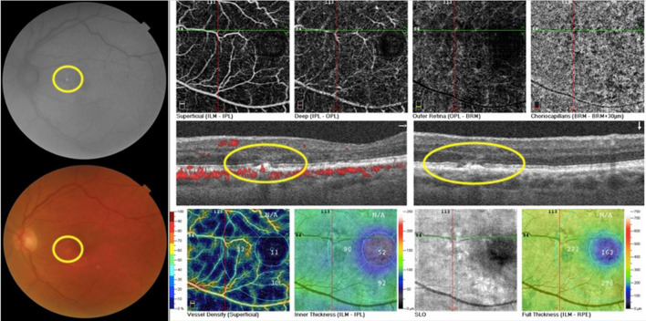
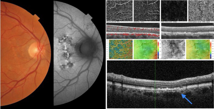
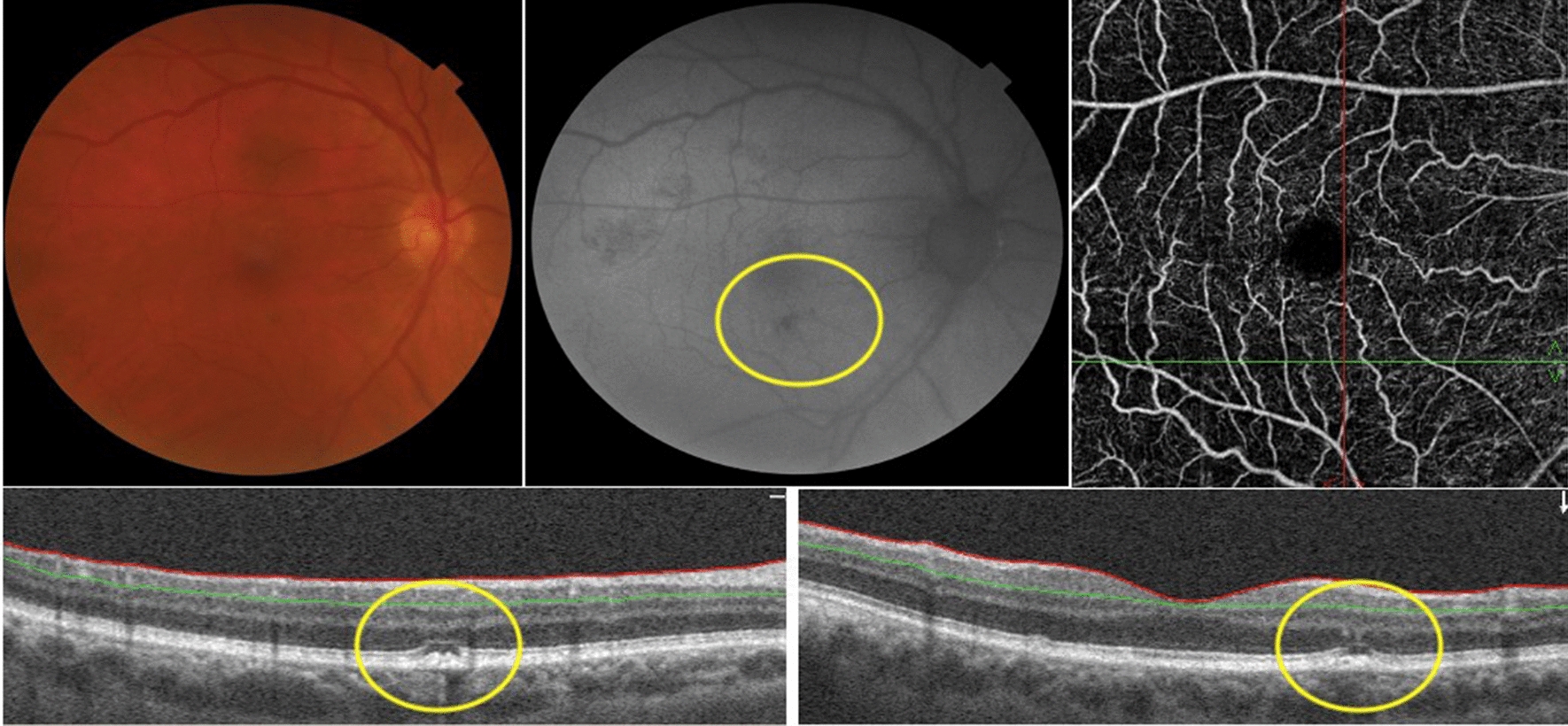
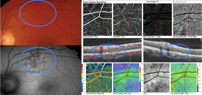
The main purpose of this study was to investigate the presence of retinal autofluorescence findings in COVID-19 patients. Observational study conducted in São Paulo in 2020. Demographic, medical history, and concomitant events, as well as medications used, hospitalization details, and laboratory test results, were obtained. Patients underwent eye examination and multimodal imaging, including color, red-free, autofluorescence fundus photography and optical coherence tomography. Eighteen patients had autofluorescence findings (6 females; average age 54 years, range 31 to 86 years; 26 eyes). Hyper-autofluorescence findings were present in 6 patients, Hypo-autofluorescence in 14 patients, and 6 patients had mixed pattern lesions. Retinal autofluorescence abnormalities were present in COVID-19 patients and may be secondary to primary or secondary changes caused by the SARS-CoV-2.

摘要

本研究的主要目的是调查新冠病毒肺炎(COVID-19)患者中视网膜自发荧光的表现。2020年在圣保罗进行了一项观察性研究。收集了患者的人口统计学信息、病史、伴随事件、用药情况、住院详情及实验室检查结果。患者接受了眼科检查和多模态成像,包括彩色、无赤光、自发荧光眼底照相及光学相干断层扫描。18例患者有自发荧光表现(6例女性;平均年龄54岁,范围31至86岁;26只眼)。6例患者出现高自发荧光表现,14例患者出现低自发荧光表现,6例患者有混合性病变。COVID-19患者存在视网膜自发荧光异常,可能继发于严重急性呼吸综合征冠状病毒2(SARS-CoV-2)引起的原发性或继发性改变。

https://cdn.ncbi.nlm.nih.gov/pmc/blobs/3b33/8626884/287d1be1a508/40942_2021_341_Fig4_HTML.jpg
https://cdn.ncbi.nlm.nih.gov/pmc/blobs/3b33/8626884/7b72ac1d8424/40942_2021_341_Fig1_HTML.jpg
https://cdn.ncbi.nlm.nih.gov/pmc/blobs/3b33/8626884/27beac43262d/40942_2021_341_Fig2_HTML.jpg
https://cdn.ncbi.nlm.nih.gov/pmc/blobs/3b33/8626884/ce6de0cbfbf8/40942_2021_341_Fig3_HTML.jpg
https://cdn.ncbi.nlm.nih.gov/pmc/blobs/3b33/8626884/287d1be1a508/40942_2021_341_Fig4_HTML.jpg

相似文献

1
Retinal autofluorescence findings after COVID-19.新冠病毒感染后的视网膜自发荧光表现
Int J Retina Vitreous. 2021 Nov 27;7(1):71. doi: 10.1186/s40942-021-00341-5.
2
MULTIMODAL IMAGING IN VORTEX VEIN VARICES.涡静脉静脉曲张的多模态成像
Retin Cases Brief Rep. 2019;13(3):260-265. doi: 10.1097/ICB.0000000000000575.
3
Multimodal imaging of white and dark without pressure fundus lesions.无压力性眼底病变的白色和黑色病变的多模态成像。
Retina. 2014 Dec;34(12):2376-87. doi: 10.1097/IAE.0000000000000388.
4
Novel optical coherence tomography classification of torpedo maculopathy.鱼雷样黄斑病变的新型光学相干断层扫描分类
Clin Exp Ophthalmol. 2015 May-Jun;43(4):342-8. doi: 10.1111/ceo.12435. Epub 2014 Nov 7.
5
Infrared fundus autofluorescence and central serous chorioretinopathy.红外眼底自发荧光与中心性浆液性脉络膜视网膜病变
Invest Ophthalmol Vis Sci. 2010 Oct;51(10):4956-62. doi: 10.1167/iovs.09-5009. Epub 2010 Apr 30.
6
Multimodal imaging characteristics of congenital grouped hyper- and hypo-pigmented fundus lesions.先天性聚集性色素增多和色素减少眼底病变的多模态影像学特征。
Clin Exp Optom. 2020 Sep;103(5):641-647. doi: 10.1111/cxo.12984. Epub 2019 Nov 25.
7
Ultra-Widefield Autofluorescence Imaging of Retinal Detachment Compared to Retinoschisis.视网膜脱离与视网膜劈裂的超广角自发荧光成像比较
Ophthalmic Surg Lasers Imaging Retina. 2019 Sep 1;50(9):550-556. doi: 10.3928/23258160-20190905-03.
8
Diabetic retinal pigment epitheliopathy: fundus autofluorescence and spectral-domain optical coherence tomography findings.糖尿病性视网膜色素上皮病变:眼底自发荧光及光谱域光学相干断层扫描结果
Graefes Arch Clin Exp Ophthalmol. 2016 Oct;254(10):1931-1940. doi: 10.1007/s00417-016-3336-8. Epub 2016 Apr 6.
9
ACUTE ZONAL OCCULT OUTER RETINOPATHY: Structural and Functional Analysis Across the Transition Zone Between Healthy and Diseased Retina.急性区域性隐匿性外层视网膜病变:健康与病变视网膜过渡区的结构与功能分析
Retina. 2018 Jan;38(1):118-127. doi: 10.1097/IAE.0000000000001513.
10
Fundus autofluorescence in type 2 idiopathic macular telangiectasia: correlation with optical coherence tomography and microperimetry.2型特发性黄斑毛细血管扩张症的眼底自发荧光:与光学相干断层扫描及微视野检查的相关性
Am J Ophthalmol. 2009 Oct;148(4):573-83. doi: 10.1016/j.ajo.2009.04.030. Epub 2009 Jul 2.

引用本文的文献

1
COVID-19 - multisystem disease.新冠病毒病——多系统疾病。
Rom J Morphol Embryol. 2025 Jan-Mar;66(1):61-67. doi: 10.47162/RJME.66.1.05.
2
The spectrum of COVID-19-associated chorioretinal vasculopathy.新型冠状病毒肺炎相关脉络膜视网膜血管病变的谱系
Am J Ophthalmol Case Rep. 2023 Sep;31:101857. doi: 10.1016/j.ajoc.2023.101857. Epub 2023 May 18.
3
Use of Imaging Technology to Assess the Effect of COVID-19 on Retinal Tissues: A Systematic Review.使用成像技术评估2019冠状病毒病对视网膜组织的影响:一项系统评价

本文引用的文献

1
Ocular findings among patients surviving COVID-19.COVID-19 幸存者的眼部表现。
Sci Rep. 2021 May 26;11(1):11085. doi: 10.1038/s41598-021-90482-2.
2
Unilateral acute posterior multifocal placoid pigment epitheliopathy in a convalescent COVID-19 patient.一名新冠康复患者的单侧急性后极部多灶性鳞状色素上皮病变
Int J Retina Vitreous. 2021 May 25;7(1):41. doi: 10.1186/s40942-021-00312-w.
3
Branch Retinal Vein Occlusion in a COVID-19 Positive Patient.一名新冠病毒检测呈阳性患者的视网膜静脉分支阻塞
Ophthalmol Ther. 2022 Jun;11(3):1017-1030. doi: 10.1007/s40123-022-00509-8. Epub 2022 Apr 29.
Cureus. 2021 Feb 27;13(2):e13586. doi: 10.7759/cureus.13586.
4
Atypical unilateral multifocal choroiditis in a COVID-19 positive patient.一名新冠病毒检测呈阳性患者的非典型单侧多灶性脉络膜炎
Am J Ophthalmol Case Rep. 2021 Jun;22:101034. doi: 10.1016/j.ajoc.2021.101034. Epub 2021 Feb 19.
5
Reply to Editorial: Interpretation of OCT and fundus findings in COVID-19 patients in recent Lancet publication.对社论的回复:近期《柳叶刀》出版物中关于新冠病毒肺炎患者光学相干断层扫描及眼底检查结果的解读
Eye (Lond). 2021 Dec;35(12):3442-3444. doi: 10.1038/s41433-020-01283-2. Epub 2020 Nov 25.
6
Bilateral Central Retinal Vein Occlusion in a 40-Year-Old Man with Severe Coronavirus Disease 2019 (COVID-19) Pneumonia.一名患有重症2019冠状病毒病(COVID-19)肺炎的40岁男性双侧视网膜中央静脉阻塞
Am J Case Rep. 2020 Oct 29;21:e927691. doi: 10.12659/AJCR.927691.
7
COVID-19 - A vascular disease.COVID-19 - 一种血管疾病。
Trends Cardiovasc Med. 2021 Jan;31(1):1-5. doi: 10.1016/j.tcm.2020.10.005. Epub 2020 Oct 14.
8
Retinal findings in hospitalised patients with severe COVID-19.住院严重 COVID-19 患者的视网膜表现。
Br J Ophthalmol. 2022 Jan;106(1):102-105. doi: 10.1136/bjophthalmol-2020-317576. Epub 2020 Oct 16.
9
Covid-19-Associated Retinopathy: A Case Report.Covid-19 相关视网膜病变:病例报告。
Ocul Immunol Inflamm. 2020 Nov 16;28(8):1293-1297. doi: 10.1080/09273948.2020.1825751. Epub 2020 Oct 6.
10
Vitritis and Outer Retinal Abnormalities in a Patient with COVID-19.COVID-19 患者的虹膜炎和外视网膜异常。
Ocul Immunol Inflamm. 2020 Nov 16;28(8):1298-1300. doi: 10.1080/09273948.2020.1821898. Epub 2020 Oct 6.