Salem Rania M, Zhang Chang, Chou Laisheng
Department of Restorative Sciences & Biomaterials, Goldman School of Dental Medicine, Boston University, Boston, MA 02118, USA.
Department of Endodontics, Goldman School of Dental Medicine, Boston University, Boston, MA 02118, USA.
Int J Biomater. 2021 Nov 18;2021:6567455. doi: 10.1155/2021/6567455. eCollection 2021.
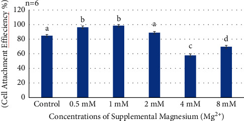
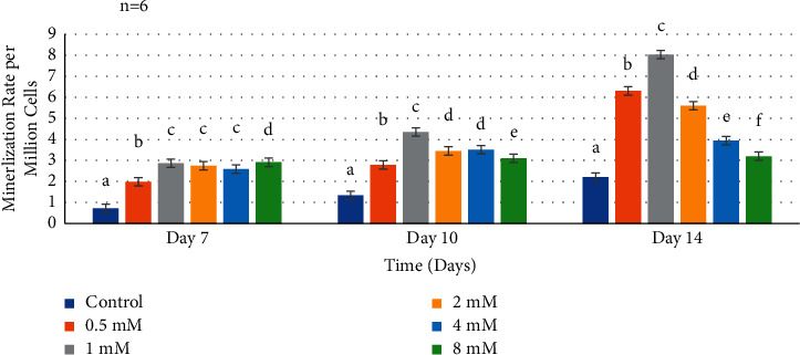
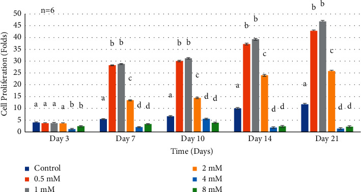
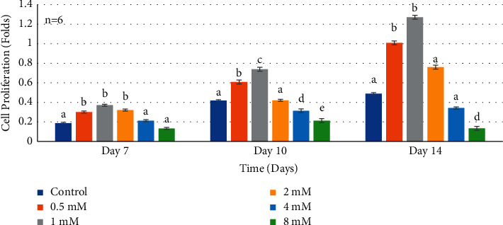
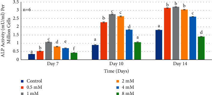
Introducing therapeutic ions into pulp capping materials has been considered a new approach for enhancing regeneration of dental tissues. However, no studies have been reported on its dentinogenic effects on human dental pulp cells (HDPCs). This study was designed to investigate the effects of magnesium (Mg) on cell attachment efficiency, proliferation, differentiation, and mineralization of HDPCs. HDPCs were cultured with 0.5 mM, 1 mM, 2 mM, 4 mM, and 8 mM concentrations of supplemental Mg and 0 mM (control). Cell attachment was measured at 4, 8, 12, 16, and 20 hours. Cell proliferation rate was evaluated at 3, 7, 10, 14, and 21 days. Crystal violet staining was used to determine cell attachment and proliferation rate. Alkaline phosphatase (ALP) activity was assessed using the fluorometric assay at 7, 10, and 14 days. Mineralization of cultures was measured by Alizarin red staining. Statistical analysis was done using multiway analysis of variance (multiway ANOVA) with Wilks' lambda test. Higher cell attachment was shown with 0.5 mM and 1 mM at 16 hours compared to control ( < 0.0001). Cells with 0.5 mM and 1 mM supplemental Mg showed significantly higher proliferation rates than control at 7, 10, 14, and 21 days ( < 0.0001). However, cell proliferation rates decreased significantly with 4 mM and 8 mM supplemental Mg at 14 and 21 days ( < 0.0001). Significantly higher levels of ALP activity and mineralization were observed in 0.5 mM, 1 mM, and 2 mM supplemental Mg at 10 and 14 days ( < 0.0001). However, 8 mM supplemental Mg showed lower ALP activity compared to control at 14 days ( < 0.0001), while 4 mM and 8 mM supplemental Mgshowed less mineralization compared to control ( < 0.0001). The study indicated that the optimal (0.5-2 mM) supplemental Mg concentrations significantly upregulated HDPCs by enhancing cell attachment, proliferation rate, ALP activity, and mineralization. Magnesium-containing biomaterials could be considered for a future novel dental pulp-capping additive in regenerative endodontics.
将治疗性离子引入牙髓盖髓材料被认为是一种促进牙组织再生的新方法。然而,尚未有关于其对人牙髓细胞(HDPCs)牙本质生成作用的研究报道。本研究旨在探讨镁(Mg)对HDPCs细胞附着效率、增殖、分化和矿化的影响。将HDPCs分别与0.5 mM、1 mM、2 mM、4 mM和8 mM浓度的补充镁以及0 mM(对照)进行培养。在4、8、12、16和20小时测量细胞附着情况。在3、7、10、14和21天评估细胞增殖率。采用结晶紫染色法测定细胞附着和增殖率。在7、10和14天使用荧光测定法评估碱性磷酸酶(ALP)活性。通过茜素红染色测量培养物的矿化情况。使用带有威尔克斯'λ检验的多因素方差分析(多因素ANOVA)进行统计分析。与对照相比,在16小时时,0.5 mM和1 mM的镁显示出更高的细胞附着(<0.0001)。在7、10、14和21天,补充0.5 mM和1 mM镁的细胞显示出比对照显著更高的增殖率(<0.0001)。然而,在14和21天,补充4 mM和8 mM镁时细胞增殖率显著下降(<0.0001)。在10和14天,在补充0.5 mM、1 mM和2 mM镁的情况下观察到显著更高水平的ALP活性和矿化(<0.0001)。然而,在14天时,8 mM补充镁与对照相比显示出较低的ALP活性(<0.0001),而4 mM和8 mM补充镁与对照相比显示出较少的矿化(<0.0001)。该研究表明,最佳(0.5 - 2 mM)补充镁浓度通过增强细胞附着、增殖率、ALP活性和矿化显著上调HDPCs。含镁生物材料可被视为未来再生牙髓病学中一种新型的牙髓盖髓添加剂。