• 文献检索
  • 文档翻译
  • 深度研究
  • 学术资讯
  • Suppr Zotero 插件Zotero 插件
  • 邀请有礼
  • 套餐&价格
  • 历史记录
应用&插件
Suppr Zotero 插件Zotero 插件浏览器插件Mac 客户端Windows 客户端微信小程序
定价
高级版会员购买积分包购买API积分包
服务
文献检索文档翻译深度研究API 文档MCP 服务
关于我们
关于 Suppr公司介绍联系我们用户协议隐私条款
关注我们

Suppr 超能文献

核心技术专利:CN118964589B侵权必究
粤ICP备2023148730 号-1Suppr @ 2026

文献检索

告别复杂PubMed语法,用中文像聊天一样搜索,搜遍4000万医学文献。AI智能推荐,让科研检索更轻松。

立即免费搜索

文件翻译

保留排版,准确专业,支持PDF/Word/PPT等文件格式,支持 12+语言互译。

免费翻译文档

深度研究

AI帮你快速写综述,25分钟生成高质量综述,智能提取关键信息,辅助科研写作。

立即免费体验

头颈癌磁共振成像中的强度标准化方法

Intensity standardization methods in magnetic resonance imaging of head and neck cancer.

作者信息

Wahid Kareem A, He Renjie, McDonald Brigid A, Anderson Brian M, Salzillo Travis, Mulder Sam, Wang Jarey, Sharafi Christina Setareh, McCoy Lance A, Naser Mohamed A, Ahmed Sara, Sanders Keith L, Mohamed Abdallah S R, Ding Yao, Wang Jihong, Hutcheson Kate, Lai Stephen Y, Fuller Clifton D, van Dijk Lisanne V

机构信息

Departments of Radiation Oncology, The University of Texas MD Anderson Cancer Center, Houston, TX, United States.

Imaging Physics, The University of Texas MD Anderson Cancer Center, Houston, TX, United States.

出版信息

Phys Imaging Radiat Oncol. 2021 Nov 20;20:88-93. doi: 10.1016/j.phro.2021.11.001. eCollection 2021 Oct.

DOI:10.1016/j.phro.2021.11.001
PMID:34849414
原文链接:https://pmc.ncbi.nlm.nih.gov/articles/PMC8607477/
Abstract

BACKGROUND AND PURPOSE

Conventional magnetic resonance imaging (MRI) poses challenges in quantitative analysis because voxel intensity values lack physical meaning. While intensity standardization methods exist, their effects on head and neck MRI have not been investigated. We developed a workflow based on healthy tissue region of interest (ROI) analysis to determine intensity consistency within a patient cohort. Through this workflow, we systematically evaluated intensity standardization methods for MRI of head and neck cancer (HNC) patients.

MATERIALS AND METHODS

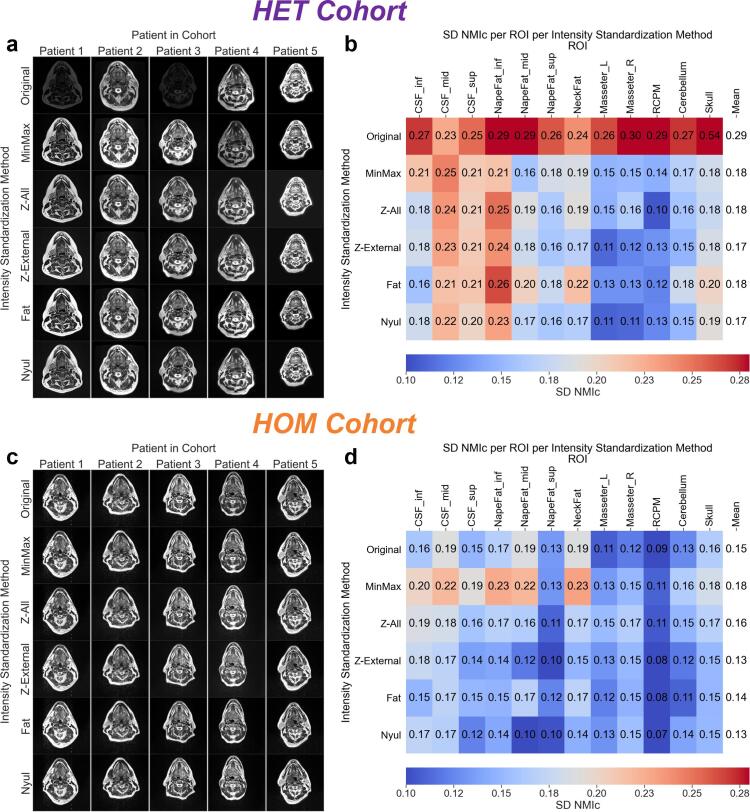
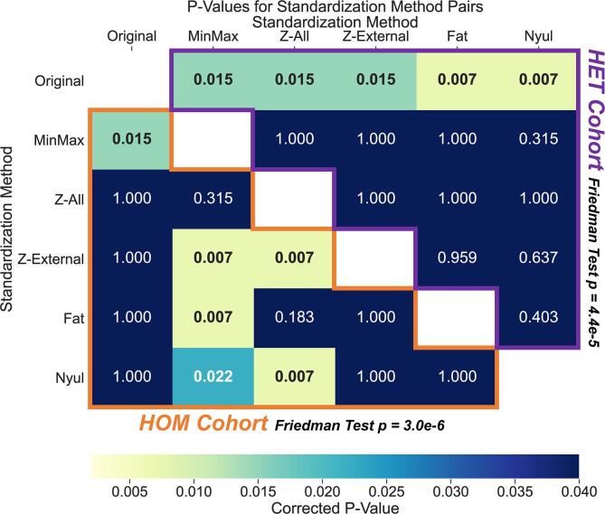
Two HNC cohorts (30 patients total) were retrospectively analyzed. One cohort was imaged with heterogenous acquisition parameters (HET cohort), whereas the other was imaged with homogenous acquisition parameters (HOM cohort). The standard deviation of cohort-level normalized mean intensity (SD NMI), a metric of intensity consistency, was calculated across ROIs to determine the effect of five intensity standardization methods on T2-weighted images. For each cohort, a Friedman test followed by a post-hoc Bonferroni-corrected Wilcoxon signed-rank test was conducted to compare SD NMI among methods.

RESULTS

Consistency (SD NMI across ROIs) between unstandardized images was substantially more impaired in the HET cohort (0.29 ± 0.08) than in the HOM cohort (0.15 ± 0.03). Consequently, corrected p-values for intensity standardization methods with lower SD NMI compared to unstandardized images were significant in the HET cohort (p < 0.05) but not significant in the HOM cohort (p > 0.05). In both cohorts, differences between methods were often minimal and nonsignificant.

CONCLUSIONS

Our findings stress the importance of intensity standardization, either through the utilization of uniform acquisition parameters or specific intensity standardization methods, and the need for testing intensity consistency before performing quantitative analysis of HNC MRI.

摘要

背景与目的

传统磁共振成像(MRI)在定量分析方面存在挑战,因为体素强度值缺乏物理意义。虽然存在强度标准化方法,但它们对头颈部MRI的影响尚未得到研究。我们开发了一种基于健康组织感兴趣区域(ROI)分析的工作流程,以确定患者队列中的强度一致性。通过此工作流程,我们系统地评估了头颈部癌(HNC)患者MRI的强度标准化方法。

材料与方法

回顾性分析了两个HNC队列(共30例患者)。一个队列采用异质采集参数成像(HET队列),而另一个队列采用同质采集参数成像(HOM队列)。计算整个ROI的队列水平归一化平均强度标准差(SD NMI),这是一种强度一致性指标,以确定五种强度标准化方法对T2加权图像的影响。对于每个队列,进行Friedman检验,然后进行事后Bonferroni校正的Wilcoxon符号秩检验,以比较各方法之间的SD NMI。

结果

未标准化图像之间的一致性(整个ROI的SD NMI)在HET队列(0.29±0.08)中比在HOM队列(0.15±0.03)中受损程度大得多。因此,与未标准化图像相比,SD NMI较低的强度标准化方法的校正p值在HET队列中具有统计学意义(p<0.05),而在HOM队列中无统计学意义(p>0.05)。在两个队列中,各方法之间的差异通常很小且无统计学意义。

结论

我们的研究结果强调了通过使用统一采集参数或特定强度标准化方法进行强度标准化的重要性,以及在对头颈部癌MRI进行定量分析之前测试强度一致性的必要性。

https://cdn.ncbi.nlm.nih.gov/pmc/blobs/b4a6/8607477/98fc586e88ab/gr2.jpg
https://cdn.ncbi.nlm.nih.gov/pmc/blobs/b4a6/8607477/c065aba0e3d1/gr1.jpg
https://cdn.ncbi.nlm.nih.gov/pmc/blobs/b4a6/8607477/98fc586e88ab/gr2.jpg
https://cdn.ncbi.nlm.nih.gov/pmc/blobs/b4a6/8607477/c065aba0e3d1/gr1.jpg
https://cdn.ncbi.nlm.nih.gov/pmc/blobs/b4a6/8607477/98fc586e88ab/gr2.jpg

相似文献

1
Intensity standardization methods in magnetic resonance imaging of head and neck cancer.头颈癌磁共振成像中的强度标准化方法
Phys Imaging Radiat Oncol. 2021 Nov 20;20:88-93. doi: 10.1016/j.phro.2021.11.001. eCollection 2021 Oct.
2
Quality assurance assessment of diagnostic and radiation therapy-simulation CT image registration for head and neck radiation therapy: anatomic region of interest-based comparison of rigid and deformable algorithms.头颈部放射治疗中诊断性CT与放射治疗模拟CT图像配准的质量保证评估:基于感兴趣解剖区域的刚性与可变形算法比较
Radiology. 2015 Mar;274(3):752-63. doi: 10.1148/radiol.14132871. Epub 2014 Nov 7.
3
Prospective quantitative quality assurance and deformation estimation of MRI-CT image registration in simulation of head and neck radiotherapy patients.对头颈部放疗患者模拟中MRI-CT图像配准进行前瞻性定量质量保证和变形估计。
Clin Transl Radiat Oncol. 2019 Apr 24;18:120-127. doi: 10.1016/j.ctro.2019.04.018. eCollection 2019 Sep.
4
Dynamic contrast enhanced-MRI in head and neck cancer patients: variability of the precontrast longitudinal relaxation time (T10).头部和颈部癌症患者的动态对比增强 MRI:预对比纵向弛豫时间(T10)的可变性。
Med Phys. 2010 Jun;37(6):2683-92. doi: 10.1118/1.3427487.
5
Comparison of cortico-medullary phase contrast-enhanced MDCT and T2-weighted MR imaging in the histological subtype differentiation of renal cell carcinoma: radiology-pathology correlation.皮质髓质期对比增强MDCT与T2加权MRI成像在肾细胞癌组织学亚型鉴别中的比较:放射学与病理学相关性
Pol J Radiol. 2021 Oct 15;86:e583-e593. doi: 10.5114/pjr.2021.111013. eCollection 2021.
6
Value of Visualization of the Intraparotid Facial Nerve and Parotid Duct Using a Micro Surface Coil and Three-Dimensional Reversed Fast Imaging With Steady-State Precession and Diffusion-Weighted Imaging Sequence.使用微表面线圈以及三维稳态进动反转快速成像和扩散加权成像序列可视化腮腺内面神经和腮腺导管的价值
J Craniofac Surg. 2018 Nov;29(8):e754-e757. doi: 10.1097/SCS.0000000000004704.
7
Serial T2-Weighted Magnetic Resonance Images Acquired on a 1.5 Tesla Magnetic Resonance Linear Accelerator Reveal Radiomic Feature Variation in Organs at Risk: An Exploratory Analysis of Novel Metrics of Tissue Response in Prostate Cancer.在1.5特斯拉磁共振直线加速器上获取的连续T2加权磁共振图像揭示了危及器官的放射组学特征变化:前列腺癌组织反应新指标的探索性分析。
Cureus. 2019 Apr 20;11(4):e4510. doi: 10.7759/cureus.4510.
8
Quantitative analysis of focal masses at MR imaging: a plea for standardization.磁共振成像中局灶性肿块的定量分析:呼吁标准化
Radiology. 2004 Jun;231(3):737-44. doi: 10.1148/radiol.2313030173.
9
Paradoxical signal pattern of mediastinal cysts on T2-weighted MR imaging: phantom and clinical study.纵隔囊肿在T2加权磁共振成像上的矛盾信号模式:模型与临床研究
Eur J Radiol. 2014 Jun;83(6):1016-1021. doi: 10.1016/j.ejrad.2014.03.004. Epub 2014 Mar 22.
10
Intravoxel incoherent motion diffusion-weighted imaging in head and neck squamous cell carcinoma: assessment of perfusion-related parameters compared to dynamic contrast-enhanced MRI.头颈部鳞状细胞癌的体素内不相干运动扩散加权成像:与动态对比增强磁共振成像相比对灌注相关参数的评估
Magn Reson Imaging. 2014 Dec;32(10):1206-13. doi: 10.1016/j.mri.2014.08.009. Epub 2014 Aug 15.

引用本文的文献

1
"Population-level age effects on the white matter structure subserving cognitive flexibility in the human brain".人群水平的年龄对人类大脑中支持认知灵活性的白质结构的影响
bioRxiv. 2025 Jul 1:2024.08.30.610526. doi: 10.1101/2024.08.30.610526.
2
Improving the U-Net Configuration for Automated Delineation of Head and Neck Cancer on MRI.改进用于在磁共振成像(MRI)上自动勾画头颈癌的U-Net配置。
Head Neck Tumor Segm MR Guid Appl (2024). 2025;15273:230-240. doi: 10.1007/978-3-031-83274-1_18. Epub 2025 Mar 3.
3
A Comprehensive Evaluation of Radiomic Features in Normal Brain Magnetic Resonance Imaging: Investigating Robustness and Region Variations.

本文引用的文献

1
Oropharyngeal primary tumor segmentation for radiotherapy planning on magnetic resonance imaging using deep learning.基于深度学习的磁共振成像口咽原发性肿瘤分割在放射治疗计划中的应用
Phys Imaging Radiat Oncol. 2021 Jul 2;19:39-44. doi: 10.1016/j.phro.2021.06.005. eCollection 2021 Jul.
2
Radiomics Repeatability Pitfalls in a Scan-Rescan MRI Study of Glioblastoma.胶质母细胞瘤扫描-重复扫描MRI研究中的影像组学重复性陷阱
Radiol Artif Intell. 2020 Dec 16;3(1):e190199. doi: 10.1148/ryai.2020190199. eCollection 2021 Jan.
3
MR-Guided Radiotherapy for Head and Neck Cancer: Current Developments, Perspectives, and Challenges.
正常脑磁共振成像中影像组学特征的综合评估:探究稳健性和区域差异
J Med Phys. 2024 Oct-Dec;49(4):608-622. doi: 10.4103/jmp.jmp_149_24. Epub 2024 Dec 18.
4
Exploring Quantitative MRI Biomarkers of Head and Neck Post-Radiation Lymphedema and Fibrosis: Post Hoc Analysis of a Prospective Trial.探索头颈部放疗后淋巴水肿和纤维化的定量MRI生物标志物:一项前瞻性试验的事后分析
Head Neck. 2025 May;47(5):1487-1496. doi: 10.1002/hed.28062. Epub 2025 Jan 10.
5
Baseline [F]FDG PET/CT and MRI first-order breast tumor features do not improve pathological complete response prediction to neoadjuvant chemotherapy.基线 [F]FDG PET/CT 和 MRI 一级乳腺癌肿瘤特征不能改善新辅助化疗的病理完全缓解预测。
Eur J Nucl Med Mol Imaging. 2024 Oct;51(12):3709-3718. doi: 10.1007/s00259-024-06815-6. Epub 2024 Jun 26.
6
Unveiling the Intricacies: A Comprehensive Review of Magnetic Resonance Imaging (MRI) Assessment of T2-Weighted Hyperintensities in the Neuroimaging Landscape.揭示复杂性:神经影像学领域中磁共振成像(MRI)对T2加权高信号评估的全面综述
Cureus. 2024 Feb 24;16(2):e54808. doi: 10.7759/cureus.54808. eCollection 2024 Feb.
7
Imaging of human papilloma virus (HPV) related oropharynx tumour: what we know to date.人乳头瘤病毒(HPV)相关口咽肿瘤的影像学:我们目前所了解的情况。
Infect Agent Cancer. 2023 Oct 9;18(1):58. doi: 10.1186/s13027-023-00530-x.
8
A transformer-based hierarchical registration framework for multimodality deformable image registration.基于变压器的多层次配准框架,用于多模态可变形图像配准。
Comput Med Imaging Graph. 2023 Sep;108:102286. doi: 10.1016/j.compmedimag.2023.102286. Epub 2023 Aug 10.
9
Magnetic resonance imaging in naso-oropharyngeal carcinoma: role of texture analysis in the assessment of response to radiochemotherapy, a preliminary study.磁共振成像在鼻腔口咽癌中的应用:纹理分析在评估放化疗反应中的作用,一项初步研究。
Radiol Med. 2023 Jul;128(7):839-852. doi: 10.1007/s11547-023-01653-2. Epub 2023 Jun 19.
10
Multimodality radiomics analysis based on [F]FDG PET/CT imaging and multisequence MRI: application to nasopharyngeal carcinoma prognosis.基于[F]FDG PET/CT 成像和多序列 MRI 的多模态放射组学分析:在鼻咽癌预后中的应用。
Eur Radiol. 2023 Oct;33(10):6677-6688. doi: 10.1007/s00330-023-09606-z. Epub 2023 Apr 15.
磁共振引导的头颈癌放疗:当前进展、前景与挑战
Front Oncol. 2021 Mar 19;11:616156. doi: 10.3389/fonc.2021.616156. eCollection 2021.
4
Simple Python Module for Conversions Between DICOM Images and Radiation Therapy Structures, Masks, and Prediction Arrays.用于 DICOM 图像与放射治疗结构、面罩和预测数组之间转换的简单 Python 模块。
Pract Radiat Oncol. 2021 May-Jun;11(3):226-229. doi: 10.1016/j.prro.2021.02.003. Epub 2021 Feb 17.
5
Magnetic resonance imaging-derived radiomic signature predicts locoregional failure after organ preservation therapy in patients with hypopharyngeal squamous cell carcinoma.磁共振成像衍生的放射组学特征可预测下咽鳞状细胞癌患者接受器官保留治疗后的局部区域复发。
Clin Transl Radiat Oncol. 2020 Aug 31;25:1-9. doi: 10.1016/j.ctro.2020.08.004. eCollection 2020 Nov.
6
Clinical variables and magnetic resonance imaging-based radiomics predict human papillomavirus status of oropharyngeal cancer.临床变量和基于磁共振成像的放射组学预测口咽癌的人乳头瘤病毒状态。
Head Neck. 2021 Feb;43(2):485-495. doi: 10.1002/hed.26505. Epub 2020 Oct 7.
7
Radiomics in medical imaging-"how-to" guide and critical reflection.医学影像中的放射组学——“操作指南”与批判性思考
Insights Imaging. 2020 Aug 12;11(1):91. doi: 10.1186/s13244-020-00887-2.
8
Standardization of brain MR images across machines and protocols: bridging the gap for MRI-based radiomics.脑磁共振图像在机器和协议间的标准化:基于 MRI 的放射组学的桥梁。
Sci Rep. 2020 Jul 23;10(1):12340. doi: 10.1038/s41598-020-69298-z.
9
Outcome prediction of head and neck squamous cell carcinoma by MRI radiomic signatures.基于 MRI 影像组学特征对头颈部鳞状细胞癌的预后预测。
Eur Radiol. 2020 Nov;30(11):6311-6321. doi: 10.1007/s00330-020-06962-y. Epub 2020 Jun 4.
10
Dynamic contrast-enhanced MRI in oncology: how we do it.动态对比增强磁共振成像在肿瘤学中的应用:我们的实践方法。
Radiol Med. 2020 Dec;125(12):1288-1300. doi: 10.1007/s11547-020-01220-z. Epub 2020 May 15.