• 文献检索
  • 文档翻译
  • 深度研究
  • 学术资讯
  • Suppr Zotero 插件Zotero 插件
  • 邀请有礼
  • 套餐&价格
  • 历史记录
应用&插件
Suppr Zotero 插件Zotero 插件浏览器插件Mac 客户端Windows 客户端微信小程序
定价
高级版会员购买积分包购买API积分包
服务
文献检索文档翻译深度研究API 文档MCP 服务
关于我们
关于 Suppr公司介绍联系我们用户协议隐私条款
关注我们

Suppr 超能文献

核心技术专利:CN118964589B侵权必究
粤ICP备2023148730 号-1Suppr @ 2026

文献检索

告别复杂PubMed语法,用中文像聊天一样搜索,搜遍4000万医学文献。AI智能推荐,让科研检索更轻松。

立即免费搜索

文件翻译

保留排版,准确专业,支持PDF/Word/PPT等文件格式,支持 12+语言互译。

免费翻译文档

深度研究

AI帮你快速写综述,25分钟生成高质量综述,智能提取关键信息,辅助科研写作。

立即免费体验

磁共振成像衍生的放射组学特征可预测下咽鳞状细胞癌患者接受器官保留治疗后的局部区域复发。

Magnetic resonance imaging-derived radiomic signature predicts locoregional failure after organ preservation therapy in patients with hypopharyngeal squamous cell carcinoma.

作者信息

Hsu Che-Yu, Lin Shih-Min, Ming Tsang Ngan, Juan Yu-Hsiang, Wang Chun-Wei, Wang Wei-Chung, Kuo Sung-Hsin

机构信息

Division of Radiation Oncology, Department of Oncology, National Taiwan University Hospital, Taipei, Taiwan.

Department of Radiation Oncology, National Taiwan University Cancer Center, National Taiwan University College of Medicine, Taipei, Taiwan.

出版信息

Clin Transl Radiat Oncol. 2020 Aug 31;25:1-9. doi: 10.1016/j.ctro.2020.08.004. eCollection 2020 Nov.

DOI:10.1016/j.ctro.2020.08.004
PMID:33426314
原文链接:https://pmc.ncbi.nlm.nih.gov/articles/PMC7780126/
Abstract

BACKGROUND AND PURPOSE

To develop and validate a magnetic resonance imaging (MRI)-derived radiomic signature (RS) for the prediction of 1-year locoregional failure (LRF) in patients with hypopharyngeal squamous cell carcinoma (HPSCC) who received organ preservation therapy (OPT).

MATERIAL AND METHODS

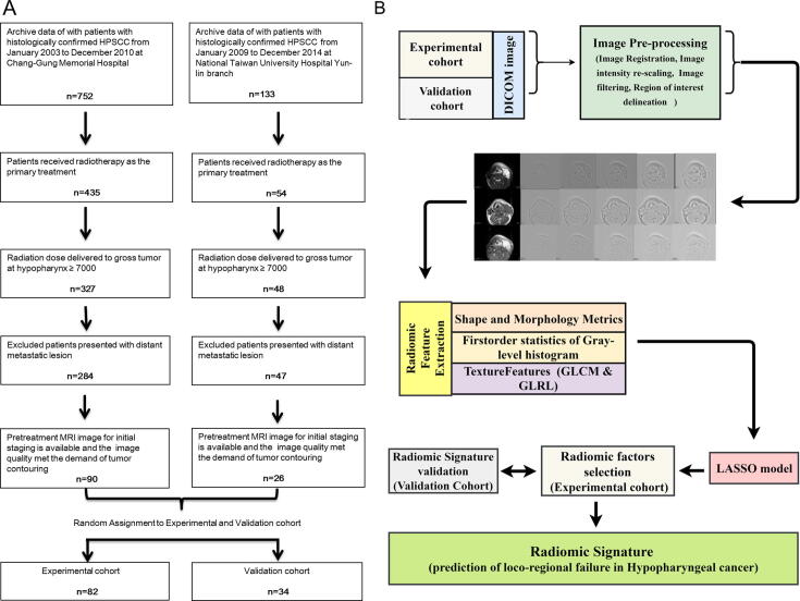
A total of 800 MRI-based features of pretreatment tumors were obtained from 116 patients with HPSCC who received OPT from two independent cohorts. The least absolute shrinkage and selection operator regression model were used to select the features used to develop the RS. Harrell's C-index and corrected C-index were used to evaluate the discriminative ability of RS. The Youden index was used to select the optimal cut-point for risk category.

RESULTS

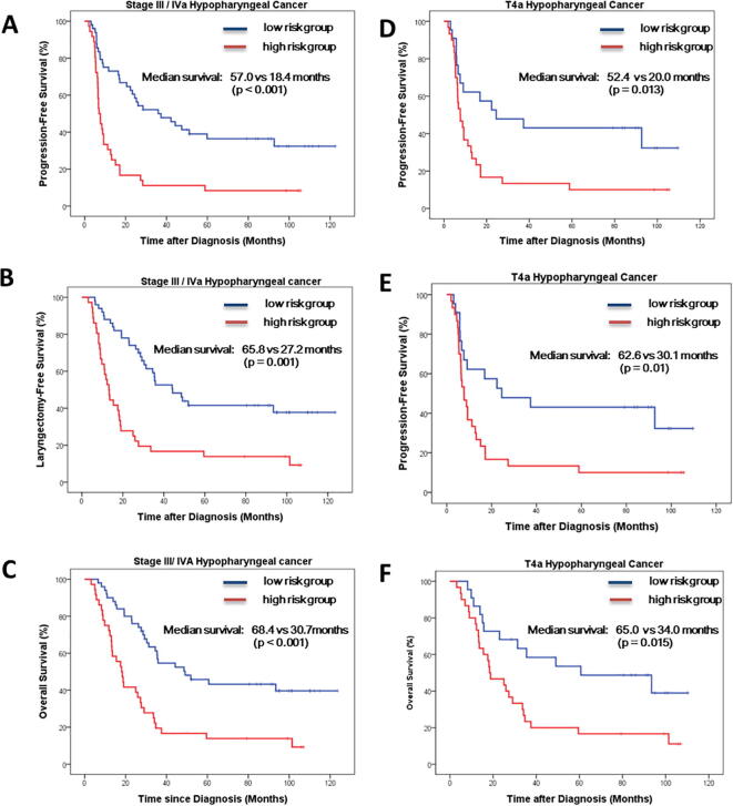
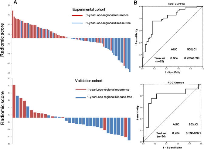
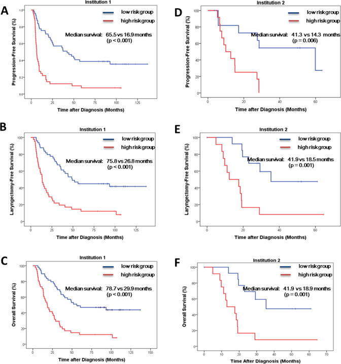
The RS yielded 1000 times bootstrapping corrected C-index of 0.8036 and 0.78235 in the experimental (n = 82) and validation cohorts (n = 34), respectively. With respect to the subgroup of patients with stage III/IV and cT4 disease, the RS also showed good predictive performance with corrected C-indices of 0.760 and 0.754, respectively. The dichotomized risk category using an RS of 0.0326 as the cut-off value yielded a 1-year LRF predictive accuracy of 79.27%, 79.41%, 76.74%, and 71.15% in the experimental, validation, stage III/IV, and cT4a cohorts, respectively. The low-risk group was associated with a significantly better progression-free laryngectomy-free and overall survival outcome in two independent institutions, stage III/IV, and cT4a cohorts.

CONCLUSION

The RS-based model provides a novel and convenient approach for the prediction of the 1-year LRF and survival outcome in patients with HPSCC who received OPT.

摘要

背景与目的

开发并验证一种基于磁共振成像(MRI)的放射组学特征(RS),用于预测接受器官保留治疗(OPT)的下咽鳞状细胞癌(HPSCC)患者的1年局部区域复发(LRF)情况。

材料与方法

从两个独立队列中接受OPT的116例HPSCC患者的预处理肿瘤中获取了总共800个基于MRI的特征。使用最小绝对收缩和选择算子回归模型选择用于开发RS的特征。采用Harrell's C指数和校正C指数评估RS的判别能力。使用约登指数选择风险分类的最佳切点。

结果

在实验队列(n = 82)和验证队列(n = 34)中,RS的1000次自举校正C指数分别为0.8036和0.78235。对于III/IV期和cT4疾病患者亚组,RS也显示出良好的预测性能,校正C指数分别为0.760和0.754。以0.0326的RS作为截断值进行二分风险分类,在实验、验证、III/IV期和cT4a队列中,1年LRF预测准确率分别为79.27%、79.41%、76.74%和71.15%。在两个独立机构、III/IV期和cT4a队列中,低风险组的无进展、无喉切除生存率和总生存结果明显更好。

结论

基于RS的模型为预测接受OPT的HPSCC患者的1年LRF和生存结果提供了一种新颖且便捷的方法。

https://cdn.ncbi.nlm.nih.gov/pmc/blobs/2254/7780126/981034e3d77c/fx1.jpg
https://cdn.ncbi.nlm.nih.gov/pmc/blobs/2254/7780126/b947228074f9/gr1.jpg
https://cdn.ncbi.nlm.nih.gov/pmc/blobs/2254/7780126/5aee1ba395fb/gr2.jpg
https://cdn.ncbi.nlm.nih.gov/pmc/blobs/2254/7780126/de0452ab3099/gr3.jpg
https://cdn.ncbi.nlm.nih.gov/pmc/blobs/2254/7780126/981034e3d77c/fx1.jpg
https://cdn.ncbi.nlm.nih.gov/pmc/blobs/2254/7780126/b947228074f9/gr1.jpg
https://cdn.ncbi.nlm.nih.gov/pmc/blobs/2254/7780126/5aee1ba395fb/gr2.jpg
https://cdn.ncbi.nlm.nih.gov/pmc/blobs/2254/7780126/de0452ab3099/gr3.jpg
https://cdn.ncbi.nlm.nih.gov/pmc/blobs/2254/7780126/981034e3d77c/fx1.jpg

相似文献

1
Magnetic resonance imaging-derived radiomic signature predicts locoregional failure after organ preservation therapy in patients with hypopharyngeal squamous cell carcinoma.磁共振成像衍生的放射组学特征可预测下咽鳞状细胞癌患者接受器官保留治疗后的局部区域复发。
Clin Transl Radiat Oncol. 2020 Aug 31;25:1-9. doi: 10.1016/j.ctro.2020.08.004. eCollection 2020 Nov.
2
Radiomics Nomogram for Predicting Locoregional Failure in Locally Advanced Non-small Cell Lung Cancer Treated with Definitive Chemoradiotherapy.基于影像组学的Nomogram 模型预测局部晚期非小细胞肺癌根治性放化疗后局部区域失败风险
Acad Radiol. 2022 Feb;29 Suppl 2:S53-S61. doi: 10.1016/j.acra.2020.11.018. Epub 2020 Dec 8.
3
Prognostic value of the radiomics-based model in progression-free survival of hypopharyngeal cancer treated with chemoradiation.基于放射组学模型在放化疗治疗下咽癌无进展生存期中的预后价值。
Eur Radiol. 2020 Feb;30(2):833-843. doi: 10.1007/s00330-019-06452-w. Epub 2019 Oct 30.
4
Predicting Progression-Free Survival Using MRI-Based Radiomics for Patients With Nonmetastatic Nasopharyngeal Carcinoma.基于MRI的影像组学预测非转移性鼻咽癌患者的无进展生存期
Front Oncol. 2020 May 12;10:618. doi: 10.3389/fonc.2020.00618. eCollection 2020.
5
Magnetic resonance imaging radiomics predicts preoperative axillary lymph node metastasis to support surgical decisions and is associated with tumor microenvironment in invasive breast cancer: A machine learning, multicenter study.磁共振成像放射组学预测术前腋窝淋巴结转移以支持手术决策,并与浸润性乳腺癌的肿瘤微环境相关:一项机器学习、多中心研究。
EBioMedicine. 2021 Jul;69:103460. doi: 10.1016/j.ebiom.2021.103460. Epub 2021 Jul 4.
6
Development and Validation of a Preoperative Magnetic Resonance Imaging Radiomics-Based Signature to Predict Axillary Lymph Node Metastasis and Disease-Free Survival in Patients With Early-Stage Breast Cancer.基于术前磁共振成像放射组学的signature 模型:预测早期乳腺癌患者腋窝淋巴结转移和无病生存的研究
JAMA Netw Open. 2020 Dec 1;3(12):e2028086. doi: 10.1001/jamanetworkopen.2020.28086.
7
Magnetic resonance imaging based radiomics signature for the preoperative discrimination of stage I-II and III-IV head and neck squamous cell carcinoma.基于磁共振成像的放射组学特征用于术前鉴别 I-II 期和 III-IV 期头颈部鳞状细胞癌。
Eur J Radiol. 2018 Sep;106:1-6. doi: 10.1016/j.ejrad.2018.07.002. Epub 2018 Jul 4.
8
Comprehensive prognostic modeling of locoregional recurrence after radiotherapy for patients with locoregionally advanced hypopharyngeal squamous cell carcinoma.局部晚期下咽鳞状细胞癌患者放疗后局部区域复发的综合预后模型
Front Oncol. 2023 Mar 21;13:1129918. doi: 10.3389/fonc.2023.1129918. eCollection 2023.
9
MRI-based radiomic signature as predictive marker for patients with head and neck squamous cell carcinoma.基于 MRI 的放射组学特征作为头颈部鳞状细胞癌患者的预测标志物。
Eur J Radiol. 2019 Aug;117:193-198. doi: 10.1016/j.ejrad.2019.06.019. Epub 2019 Jun 25.
10
Whole-Volume Tumor MRI Radiomics for Prognostic Modeling in Endometrial Cancer.全容积肿瘤 MRI 影像组学在子宫内膜癌预后建模中的应用。
J Magn Reson Imaging. 2021 Mar;53(3):928-937. doi: 10.1002/jmri.27444. Epub 2020 Nov 16.

引用本文的文献

1
An MRI-based radiomics nomogram for detecting cervical esophagus invasion in hypopharyngeal squamous cell carcinoma.基于 MRI 的放射组学列线图用于检测下咽鳞癌中颈段食管侵犯。
Cancer Imaging. 2023 Dec 15;23(1):120. doi: 10.1186/s40644-023-00642-y.
2
Radiomics in Hypopharyngeal Cancer Management: A State-of-the-Art Review.下咽癌管理中的影像组学:最新综述
Biomedicines. 2023 Mar 6;11(3):805. doi: 10.3390/biomedicines11030805.
3
Exploratory ensemble interpretable model for predicting local failure in head and neck cancer: the additive benefit of CT and intra-treatment cone-beam computed tomography features.

本文引用的文献

1
Prognostic value of lymph node ratio in laryngeal and hypopharyngeal squamous cell carcinoma: a systematic review and meta-analysis.淋巴结比率在喉和下咽鳞状细胞癌中的预后价值:系统评价和荟萃分析。
J Otolaryngol Head Neck Surg. 2020 May 29;49(1):31. doi: 10.1186/s40463-020-00421-w.
2
Radiomics Driven Diffusion Weighted Imaging Sensing Strategies for Zone-Level Prostate Cancer Sensing.基于放射组学的扩散加权成像传感策略在前列腺癌分区水平检测中的应用。
Sensors (Basel). 2020 Mar 10;20(5):1539. doi: 10.3390/s20051539.
3
Heterogeneity in tumours: Validating the use of radiomic features on F-FDG PET/CT scans of lung cancer patients as a prognostic tool.
用于预测头颈癌局部失败的探索性集成可解释模型:CT和治疗中锥形束计算机断层扫描特征的附加益处
Quant Imaging Med Surg. 2021 Dec;11(12):4781-4796. doi: 10.21037/qims-21-274.
4
Intensity standardization methods in magnetic resonance imaging of head and neck cancer.头颈癌磁共振成像中的强度标准化方法
Phys Imaging Radiat Oncol. 2021 Nov 20;20:88-93. doi: 10.1016/j.phro.2021.11.001. eCollection 2021 Oct.
5
An MRI-based radiomics-clinical nomogram for the overall survival prediction in patients with hypopharyngeal squamous cell carcinoma: a multi-cohort study.基于 MRI 的放射组学-临床列线图预测下咽鳞癌患者总生存:多队列研究。
Eur Radiol. 2022 Mar;32(3):1548-1557. doi: 10.1007/s00330-021-08292-z. Epub 2021 Oct 19.
肿瘤异质性:验证使用肺癌患者 F-FDG PET/CT 扫描的放射组学特征作为预后工具的有效性。
Radiother Oncol. 2020 Mar;144:72-78. doi: 10.1016/j.radonc.2019.10.012. Epub 2019 Nov 14.
4
Human papillomavirus-associated squamous cell carcinoma of the larynx or hypopharynx: Clinical outcomes and implications for laryngeal preservation.人乳头瘤病毒相关性喉或下咽鳞状细胞癌:临床结果及对保留喉功能的影响。
Oral Oncol. 2019 Nov;98:20-27. doi: 10.1016/j.oraloncology.2019.09.008. Epub 2019 Sep 16.
5
Radiomic Signatures Derived from Diffusion-Weighted Imaging for the Assessment of Breast Cancer Receptor Status and Molecular Subtypes.基于扩散加权成像的影像组学特征评估乳腺癌受体状态和分子亚型。
Mol Imaging Biol. 2020 Apr;22(2):453-461. doi: 10.1007/s11307-019-01383-w.
6
Organ preservation in laryngeal and hypopharyngeal cancer.喉癌和下咽癌的器官保存
Oral Oncol. 2019 Mar;90:6-7. doi: 10.1016/j.oraloncology.2019.01.014. Epub 2019 Jan 28.
7
The Current Indications for Non-Surgical Treatment of Hypopharyngeal Cancer.下咽癌非手术治疗的当前适应证
Adv Otorhinolaryngol. 2019;83:76-89. doi: 10.1159/000492314. Epub 2019 Feb 12.
8
NCTN Assessment on Current Applications of Radiomics in Oncology.NCTN 评估放射组学在肿瘤学中的当前应用。
Int J Radiat Oncol Biol Phys. 2019 Jun 1;104(2):302-315. doi: 10.1016/j.ijrobp.2019.01.087. Epub 2019 Jan 31.
9
CT imaging during treatment improves radiomic models for patients with locally advanced head and neck cancer.治疗期间的 CT 成像可改善局部晚期头颈部癌症患者的放射组学模型。
Radiother Oncol. 2019 Jan;130:10-17. doi: 10.1016/j.radonc.2018.07.020. Epub 2018 Aug 4.
10
The Use of Quantitative Imaging in Radiation Oncology: A Quantitative Imaging Network (QIN) Perspective.定量影像学在放射肿瘤学中的应用:定量影像学网络(QIN)视角。
Int J Radiat Oncol Biol Phys. 2018 Nov 15;102(4):1219-1235. doi: 10.1016/j.ijrobp.2018.06.023. Epub 2018 Jun 30.