Li Hao, Zhang Huahong, Johnson Hans, Long Jeffrey D, Paulsen Jane S, Oguz Ipek
Department of Electrical Engineering and Computer Science, Vanderbilt University, Nashville, TN 37235.
Department of Electrical and Computer Engineering, University of Iowa, Iowa City, IA 52242.
Proc SPIE Int Soc Opt Eng. 2021 Feb;11596. doi: 10.1117/12.2582005. Epub 2021 Feb 15.
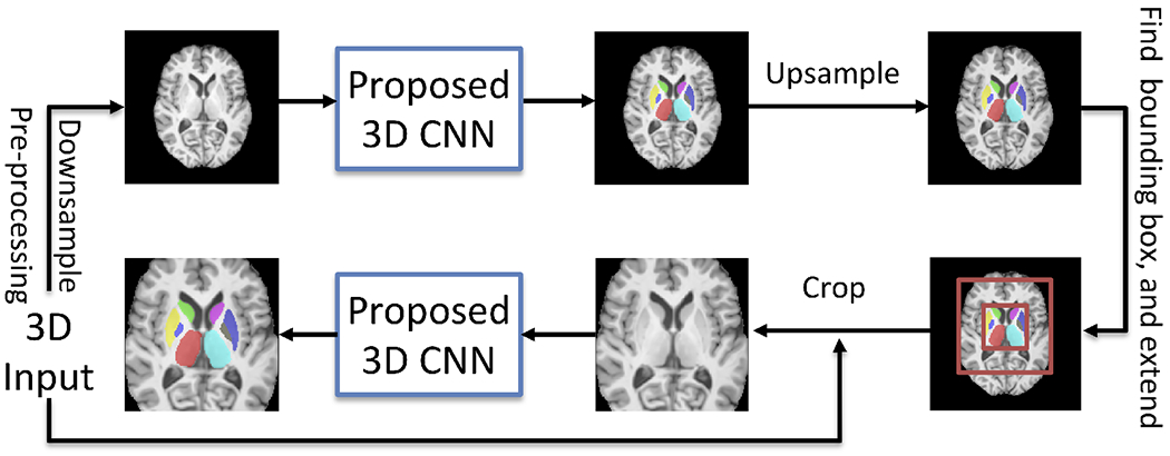
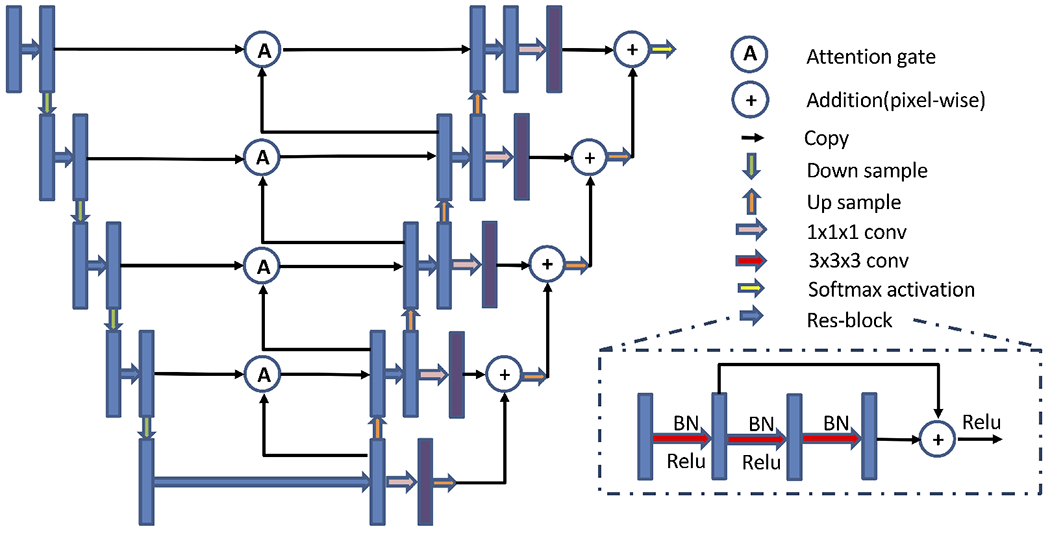
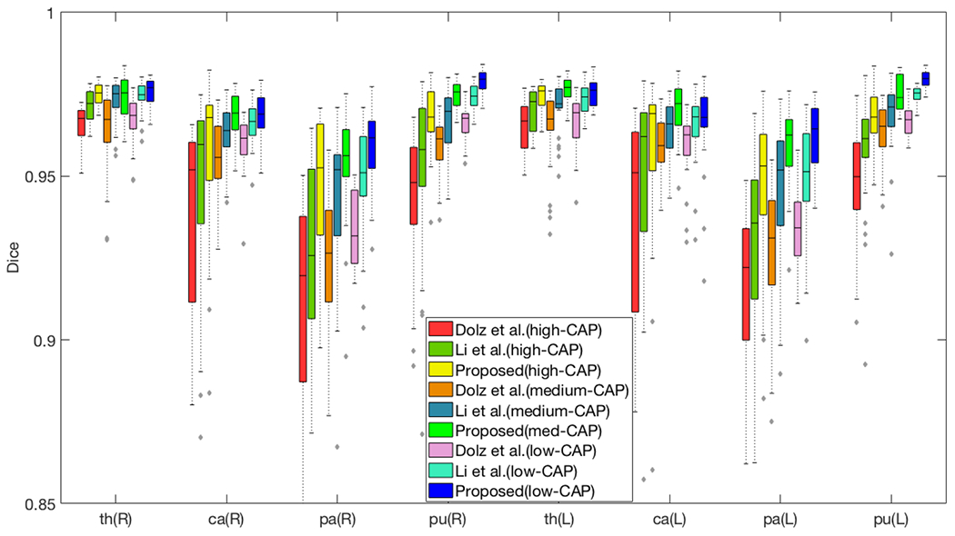
The subcortical structures of the brain are relevant for many neurodegenerative diseases like Huntington's disease (HD). Quantitative segmentation of these structures from magnetic resonance images (MRIs) has been studied in clinical and neuroimaging research. Recently, convolutional neural networks (CNNs) have been successfully used for many medical image analysis tasks, including subcortical segmentation. In this work, we propose a 2-stage cascaded 3D subcortical segmentation framework, with the same 3D CNN architecture for both stages. Attention gates, residual blocks and output adding are used in our proposed 3D CNN. In the first stage, we apply our model to downsampled images to output a coarse segmentation. Next, we crop the extended subcortical region from the original image based on this coarse segmentation, and we input the cropped region to the second CNN to obtain the final segmentation. Left and right pairs of thalamus, caudate, pallidum and putamen are considered in our segmentation. We use the Dice coefficient as our metric and evaluate our method on two datasets: the publicly available IBSR dataset and a subset of the PREDICT-HD database, which includes healthy controls and HD subjects. We train our models on only healthy control subjects and test on both healthy controls and HD subjects to examine model generalizability. Compared with the state-of-the-art methods, our method has the highest mean Dice score on all considered subcortical structures (except the thalamus on IBSR), with more pronounced improvement for HD subjects. This suggests that our method may have better ability to segment MRIs of subjects with neurodegenerative disease.
大脑的皮层下结构与许多神经退行性疾病相关,如亨廷顿舞蹈病(HD)。在临床和神经影像学研究中,已经对从磁共振图像(MRI)中对这些结构进行定量分割展开了研究。最近,卷积神经网络(CNN)已成功应用于许多医学图像分析任务,包括皮层下分割。在这项工作中,我们提出了一个两阶段级联的3D皮层下分割框架,两个阶段采用相同的3D CNN架构。我们提出的3D CNN中使用了注意力门、残差块和输出相加。在第一阶段,我们将模型应用于下采样图像以输出粗略分割。接下来,我们基于此粗略分割从原始图像中裁剪出扩展的皮层下区域,并将裁剪后的区域输入到第二个CNN中以获得最终分割。我们的分割考虑了丘脑、尾状核、苍白球和壳核的左右对。我们使用Dice系数作为度量标准,并在两个数据集上评估我们的方法:公开可用的IBSR数据集和PREDICT-HD数据库的一个子集,该子集包括健康对照和HD受试者。我们仅在健康对照受试者上训练模型,并在健康对照和HD受试者上进行测试,以检验模型的泛化能力。与现有最先进的方法相比,我们的方法在所有考虑的皮层下结构上(IBSR数据集上的丘脑除外)具有最高的平均Dice分数,对HD受试者的改善更为显著。这表明我们的方法可能具有更好的分割神经退行性疾病受试者MRI的能力。