Section of Radiology- Department of Biomedicine, Neurosciences and Advanced Diagnostics (BiND), University Hospital "Paolo Giaccone", Via del Vespro 129, 90127, Palermo, Italy.
Department of Health Promotion, Mother and Child Care, Internal Medicine and Medical Specialties (PROMISE), University Hospital of Palermo, Via del Vespro 129, 90127, Palermo, Italy.
Eur Radiol Exp. 2021 Dec 7;5(1):52. doi: 10.1186/s41747-021-00250-0.
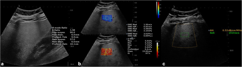
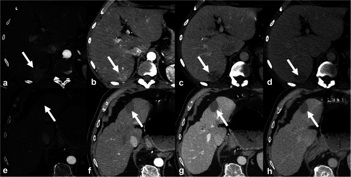
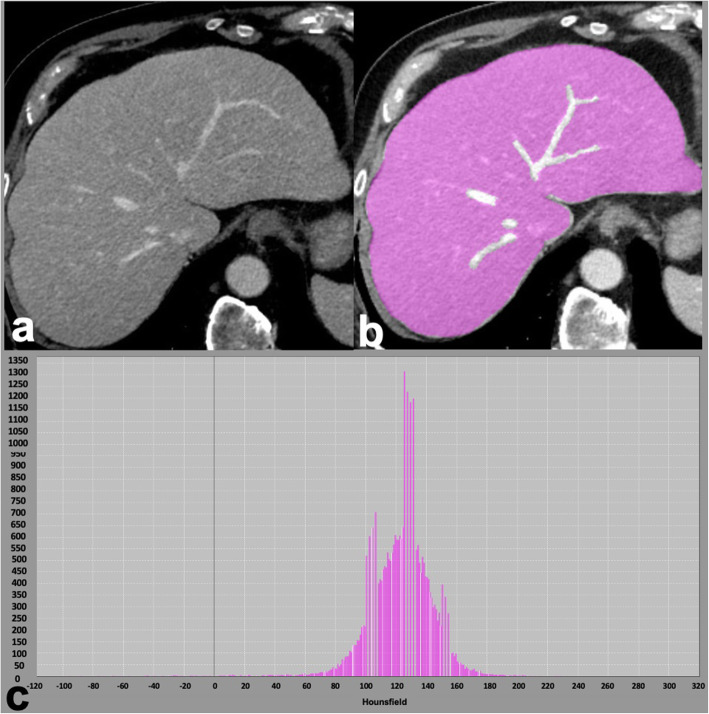
Over the past two decades, the epidemiology of chronic liver disease has changed with an increase in the prevalence of nonalcoholic fatty liver disease in parallel to the advent of curative treatments for hepatitis C. Recent developments provided new tools for diagnosis and monitoring of liver diseases based on ultrasound (US), computed tomography (CT), and magnetic resonance imaging (MRI), as applied for assessing steatosis, fibrosis, and focal lesions. This narrative review aims to discuss the emerging approaches for qualitative and quantitative liver imaging, focusing on those expected to become adopted in clinical practice in the next 5 to 10 years. While radiomics is an emerging tool for many of these applications, dedicated techniques have been investigated for US (controlled attenuation parameter, backscatter coefficient, elastography methods such as point shear wave elastography [pSWE] and transient elastography [TE], novel Doppler techniques, and three-dimensional contrast-enhanced ultrasound [3D-CEUS]), CT (dual-energy, spectral photon counting, extracellular volume fraction, perfusion, and surface nodularity), and MRI (proton density fat fraction [PDFF], elastography [MRE], contrast enhancement index, relative enhancement, T1 mapping on the hepatobiliary phase, perfusion). Concurrently, the advent of abbreviated MRI protocols will help fulfill an increasing number of examination requests in an era of healthcare resource constraints.
在过去的二十年中,随着丙型肝炎治疗方法的出现,非酒精性脂肪性肝病的患病率也在增加,慢性肝病的流行病学也发生了变化。最近的发展为超声(US)、计算机断层扫描(CT)和磁共振成像(MRI)诊断和监测肝脏疾病提供了新的工具,用于评估脂肪变性、纤维化和局灶性病变。本叙述性综述旨在讨论定性和定量肝脏成像的新方法,重点介绍预计在未来 5 到 10 年内将在临床实践中采用的方法。虽然放射组学是这些应用的一个新兴工具,但已针对 US(受控衰减参数、反向散射系数、弹性成像方法如点剪切波弹性成像[pSWE]和瞬时弹性成像[TE]、新型多普勒技术和三维对比增强超声[3D-CEUS])、CT(双能、光谱光子计数、细胞外体积分数、灌注和表面结节性)和 MRI(质子密度脂肪分数[PDFF]、弹性成像[MRE]、对比增强指数、相对增强、肝胆期 T1 映射、灌注)进行了专门技术研究。同时,缩短 MRI 方案的出现将有助于在医疗资源有限的时代满足越来越多的检查请求。