Department of Women's and Children's Health, Centre for Women's Health Research, Institute of Life Course and Medical Sciences, University of Liverpool, Member of Liverpool Health Partners, Liverpool, UK.
Liverpool Women's NHS Foundation Trust, Member of Liverpool Health Partners, Liverpool, UK.
Hum Reprod Update. 2022 Feb 28;28(2):153-171. doi: 10.1093/humupd/dmab039.
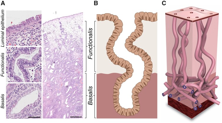
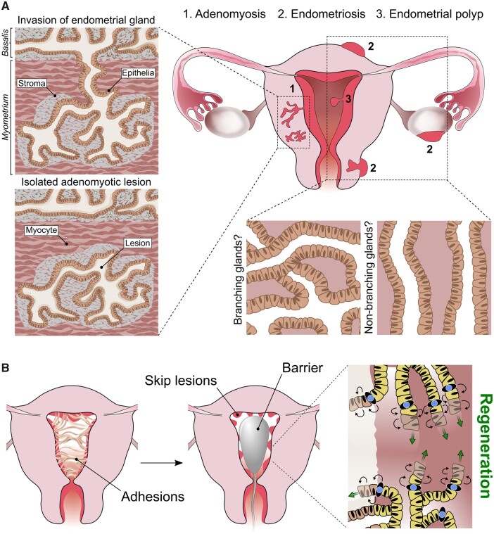
Human endometrium remains a poorly understood tissue of the female reproductive tract. The superficial endometrial functionalis, the site of embryo implantation, is repeatedly shed with menstruation, and the stem cell-rich deeper basalis is postulated to be responsible for the regeneration of the functionalis. Two recent manuscripts have demonstrated the 3D architecture of endometrial glands. These manuscripts have challenged and replaced the prevailing concept that these glands end in blind pouches in the basalis layer that contain stem cells in crypts, as in the intestinal mucosa, providing a new paradigm for endometrial glandular anatomy. This necessitates re-evaluation of the available evidence on human endometrial regeneration in both health and disease in the context of this previously unknown endometrial glandular arrangement.
The aim of this review is to determine if the recently discovered glandular arrangement provides plausible explanations for previously unanswered questions related to human endometrial biology. Specifically, it will focus on re-appraising the theories related to endometrial regeneration, location of stem/progenitor cells and endometrial pathologies in the context of this recently unravelled endometrial glandular organization.
An extensive literature search was conducted from inception to April 2021 using multiple databases, including PubMed/Web of Science/EMBASE/Scopus, to select studies using keywords applied to endometrial glandular anatomy and regeneration, and the references included in selected publications were also screened. All relevant publications were included.
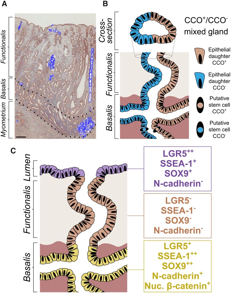
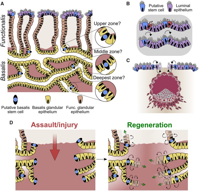
The human endometrial glands have a unique and complex architecture; branched basalis glands proceed in a horizontal course adjacent to the myometrium, as opposed to the non-branching, vertically coiled functionalis glands, which run parallel to each other as is observed in intestinal crypts. This complex network of mycelium-like, interconnected basalis glands is demonstrated to contain endometrial epithelial stem cells giving rise to single, non-branching functionalis glands. Several previous studies that have tried to confirm the existence of epithelial stem cells have used methodologies that prevent sampling of the stem cell-rich basalis. More recent findings have provided insight into the efficient regeneration of the human endometrium, which is preferentially evolved in humans and menstruating upper-order primates.
The unique physiological organization of the human endometrial glandular element, its relevance to stem cell activity and scarless endometrial regeneration will inform reproductive biologists and clinicians to direct their future research to determine disease-specific alterations in glandular anatomy in a variety of endometrial pathological conditions.
人类子宫内膜仍然是女性生殖道中一个尚未被充分了解的组织。位于子宫内膜表面的功能层会随着月经脱落,而富含干细胞的深部基底层被认为负责功能层的再生。最近有两篇论文描述了子宫内膜腺体的 3D 结构。这两篇论文挑战并取代了现有的观点,即这些腺体在基底层中以盲袋的形式结束,这些盲袋中包含位于隐窝内的干细胞,就像在肠黏膜中一样,为子宫内膜腺体的解剖结构提供了一个新的范例。这就需要在这一未知的子宫内膜腺体排列方式的背景下,重新评估现有关于健康和疾病状态下人类子宫内膜再生的证据。
本综述的目的是确定最近发现的腺体排列方式是否为与人类子宫内膜生物学相关的先前未回答的问题提供了合理的解释。具体来说,它将重点重新评估与子宫内膜再生、干细胞/祖细胞位置以及子宫内膜病变相关的理论,以了解这一新揭示的子宫内膜腺体组织的相关内容。
从 2021 年 4 月开始,使用多个数据库(包括 PubMed/Web of Science/EMBASE/Scopus)进行了广泛的文献检索,使用关键词选择与子宫内膜腺体解剖和再生相关的研究,并对选定出版物中的参考文献进行了筛选。所有相关出版物均被纳入。
人类子宫内膜腺体具有独特而复杂的结构;分支的基底层腺体呈水平方向毗邻子宫肌层,而不是像非分支的、垂直盘旋的功能层腺体那样彼此平行,这与肠隐窝中的情况相似。这种复杂的菌丝状、相互连接的基底层腺体网络被证明包含产生单个非分支功能层腺体的子宫内膜上皮干细胞。之前有几项试图证实上皮干细胞存在的研究使用了阻止采样富含干细胞的基底层的方法。最近的发现提供了对人类子宫内膜有效再生的深入了解,这是人类和有经期的高级灵长类动物特有的。
人类子宫内膜腺体独特的生理组织结构、其与干细胞活性和无瘢痕子宫内膜再生的关系,将为生殖生物学家和临床医生提供信息,指导他们未来的研究,以确定各种子宫内膜病理条件下腺体解剖结构的特定疾病变化。