• 文献检索
  • 文档翻译
  • 深度研究
  • 学术资讯
  • Suppr Zotero 插件Zotero 插件
  • 邀请有礼
  • 套餐&价格
  • 历史记录
应用&插件
Suppr Zotero 插件Zotero 插件浏览器插件Mac 客户端Windows 客户端微信小程序
定价
高级版会员购买积分包购买API积分包
服务
文献检索文档翻译深度研究API 文档MCP 服务
关于我们
关于 Suppr公司介绍联系我们用户协议隐私条款
关注我们

Suppr 超能文献

核心技术专利:CN118964589B侵权必究
粤ICP备2023148730 号-1Suppr @ 2026

文献检索

告别复杂PubMed语法,用中文像聊天一样搜索,搜遍4000万医学文献。AI智能推荐,让科研检索更轻松。

立即免费搜索

文件翻译

保留排版,准确专业,支持PDF/Word/PPT等文件格式,支持 12+语言互译。

免费翻译文档

深度研究

AI帮你快速写综述,25分钟生成高质量综述,智能提取关键信息,辅助科研写作。

立即免费体验

磁共振成像衍生的g值比率与新诊断多发性硬化症的病灶严重程度

MRI-derived g-ratio and lesion severity in newly diagnosed multiple sclerosis.

作者信息

York Elizabeth N, Martin Sarah-Jane, Meijboom Rozanna, Thrippleton Michael J, Bastin Mark E, Carter Edwin, Overell James, Connick Peter, Chandran Siddharthan, Waldman Adam D, Hunt David P J

机构信息

Centre for Clinical Brain Sciences, University of Edinburgh, Edinburgh EH16 4SB, UK.

Department of Neurosciences, University of Glasgow, Glasgow G51 4LB, UK.

出版信息

Brain Commun. 2021 Nov 3;3(4):fcab249. doi: 10.1093/braincomms/fcab249. eCollection 2021.

DOI:10.1093/braincomms/fcab249
PMID:34877533
原文链接:https://pmc.ncbi.nlm.nih.gov/articles/PMC8643503/
Abstract

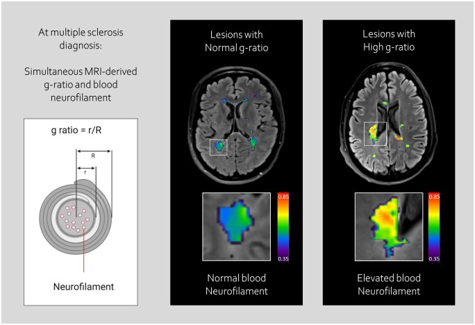
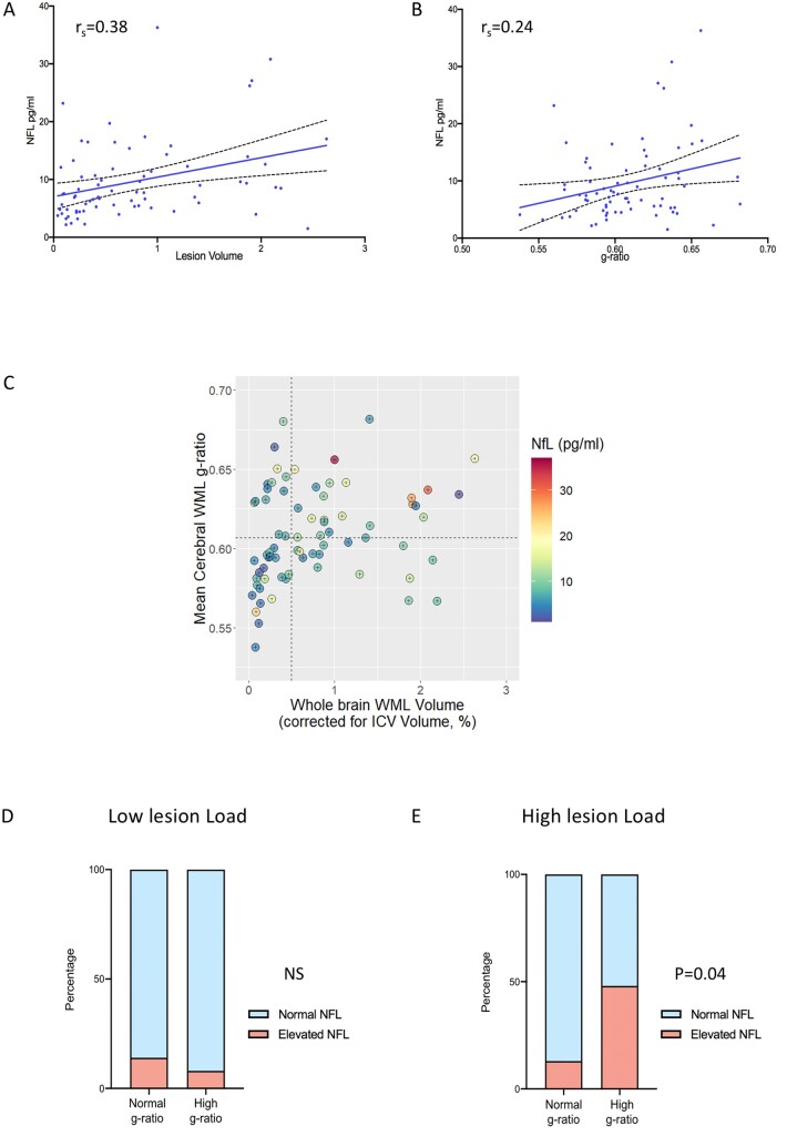
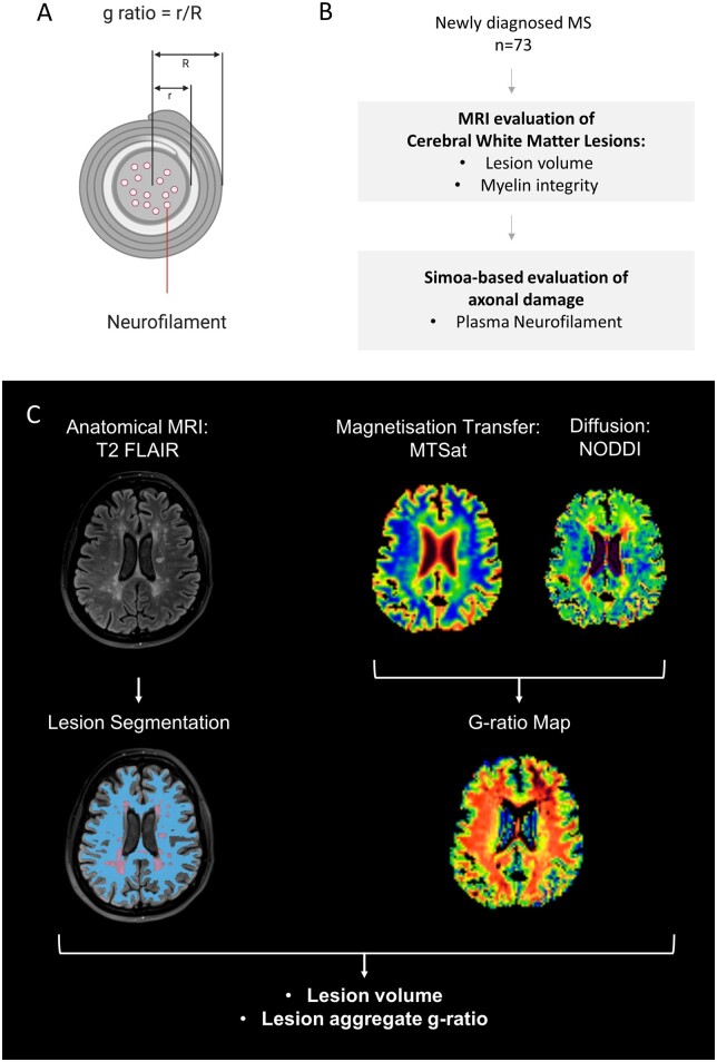
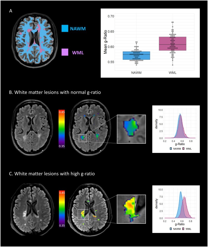
Myelin loss is associated with axonal damage in established multiple sclerosis. This relationship is challenging to study in early disease. Here, we ask whether myelin loss is associated with axonal damage at diagnosis by combining non-invasive neuroimaging and blood biomarkers. We performed quantitative microstructural MRI and single-molecule ELISA plasma neurofilament measurement in 73 patients with newly diagnosed, immunotherapy naïve relapsing-remitting multiple sclerosis. Myelin integrity was evaluated using aggregate g-ratios, derived from magnetization transfer saturation and neurite orientation dispersion and density imaging diffusion data. We found significantly higher g-ratios within cerebral white matter lesions (suggesting myelin loss) compared with normal-appearing white matter (0.61 versus 0.57, difference 0.036, 95% CI: 0.029-0.043, < 0.001). Lesion volume (Spearman's rho r= 0.38, < 0.001) and g-ratio (r= 0.24, < 0.05) correlated independently with plasma neurofilament. In patients with substantial lesion load ( = 38), those with higher g-ratio (defined as greater than median) were more likely to have abnormally elevated plasma neurofilament than those with normal g-ratio (defined as less than median) [11/23 (48%) versus 2/15 (13%), < 0.05]. These data suggest that, even at multiple sclerosis diagnosis, reduced myelin integrity is associated with axonal damage. MRI-derived g-ratio may provide useful additional information regarding lesion severity and help to identify individuals with a high degree of axonal damage at disease onset.

摘要

在已确诊的多发性硬化症中,髓鞘损失与轴突损伤相关。这种关系在疾病早期很难进行研究。在这里,我们通过结合非侵入性神经影像学和血液生物标志物来探讨在诊断时髓鞘损失是否与轴突损伤相关。我们对73例新诊断的、未接受过免疫治疗的复发缓解型多发性硬化症患者进行了定量微观结构MRI和单分子ELISA血浆神经丝测量。使用从磁化传递饱和度、神经突方向离散度和密度成像扩散数据得出的总体g比率来评估髓鞘完整性。我们发现,与外观正常的白质相比,脑白质病变内的g比率显著更高(表明髓鞘损失)(0.61对0.57,差异0.036,95%可信区间:0.029 - 0.043,<0.001)。病变体积(斯皮尔曼等级相关系数r = 0.38,<0.001)和g比率(r = 0.24,<0.05)与血浆神经丝独立相关。在病变负荷较大的患者(n = 38)中,g比率较高(定义为大于中位数)的患者比g比率正常(定义为小于中位数)的患者更有可能出现血浆神经丝异常升高[11/23(48%)对2/15(13%),<0.05]。这些数据表明,即使在多发性硬化症诊断时,髓鞘完整性降低也与轴突损伤相关。MRI得出的g比率可能为病变严重程度提供有用的额外信息,并有助于识别疾病发作时轴突损伤程度较高的个体。

https://cdn.ncbi.nlm.nih.gov/pmc/blobs/f905/8643503/b5995c95330c/fcab249f3.jpg
https://cdn.ncbi.nlm.nih.gov/pmc/blobs/f905/8643503/b365e0ccf19d/fcab249f4.jpg
https://cdn.ncbi.nlm.nih.gov/pmc/blobs/f905/8643503/775830602363/fcab249f1.jpg
https://cdn.ncbi.nlm.nih.gov/pmc/blobs/f905/8643503/08a5b465f038/fcab249f2.jpg
https://cdn.ncbi.nlm.nih.gov/pmc/blobs/f905/8643503/b5995c95330c/fcab249f3.jpg

相似文献

1
MRI-derived g-ratio and lesion severity in newly diagnosed multiple sclerosis.磁共振成像衍生的g值比率与新诊断多发性硬化症的病灶严重程度
Brain Commun. 2021 Nov 3;3(4):fcab249. doi: 10.1093/braincomms/fcab249. eCollection 2021.
2
Longitudinal microstructural MRI markers of demyelination and neurodegeneration in early relapsing-remitting multiple sclerosis: Magnetisation transfer, water diffusion and g-ratio.早期复发缓解型多发性硬化症的脱髓鞘和神经退行性变的纵向微观结构 MRI 标志物:磁化传递、水扩散和 g 比值。
Neuroimage Clin. 2022;36:103228. doi: 10.1016/j.nicl.2022.103228. Epub 2022 Oct 13.
3
Myelin and axon pathology in multiple sclerosis assessed by myelin water and multi-shell diffusion imaging.多发性硬化症的髓鞘和轴突病理通过髓鞘水和多壳扩散成像评估。
Brain. 2021 Jul 28;144(6):1684-1696. doi: 10.1093/brain/awab088.
4
In vivo characterization of cortical and white matter neuroaxonal pathology in early multiple sclerosis.早期多发性硬化症中皮质和白质神经轴突病变的体内特征分析
Brain. 2017 Nov 1;140(11):2912-2926. doi: 10.1093/brain/awx247.
5
Serum neurofilament light chain correlates with myelin and axonal magnetic resonance imaging markers in multiple sclerosis.血清神经丝轻链与多发性硬化症的髓鞘和轴索磁共振成像标志物相关。
Mult Scler Relat Disord. 2022 Jan;57:103366. doi: 10.1016/j.msard.2021.103366. Epub 2021 Nov 3.
6
Neurite orientation dispersion and density imaging in myelin oligodendrocyte glycoprotein antibody-associated disease and neuromyelitis optica spectrum disorders.髓鞘少突胶质细胞糖蛋白抗体相关疾病和视神经脊髓炎谱系障碍中的神经突方向离散度与密度成像
Mult Scler Relat Disord. 2025 Mar;95:106324. doi: 10.1016/j.msard.2025.106324. Epub 2025 Feb 8.
7
Brain microstructural and metabolic alterations detected in vivo at onset of the first demyelinating event.在首次脱髓鞘事件发作时体内检测到的脑微观结构和代谢改变。
Brain. 2021 Jun 22;144(5):1409-1421. doi: 10.1093/brain/awab043.
8
Proteomics and relationship with axonal pathology in multiple sclerosis: 5-year diffusion tensor imaging study.蛋白质组学与多发性硬化症轴突病理学的关系:5年扩散张量成像研究
Brain Commun. 2023 Jun 13;5(3):fcad183. doi: 10.1093/braincomms/fcad183. eCollection 2023.
9
Imaging G-Ratio in Multiple Sclerosis Using High-Gradient Diffusion MRI and Macromolecular Tissue Volume.使用高梯度弥散 MRI 和大分子组织容积成像技术评估多发性硬化症的 G 比值
AJNR Am J Neuroradiol. 2019 Nov;40(11):1871-1877. doi: 10.3174/ajnr.A6283. Epub 2019 Nov 6.
10
Patterns of cerebral damage in multiple sclerosis and aquaporin-4 antibody-positive neuromyelitis optica spectrum disorders-major differences revealed by non-conventional imaging.多发性硬化症和水通道蛋白4抗体阳性视神经脊髓炎谱系障碍中的脑损伤模式——非常规成像揭示的主要差异
Brain Commun. 2024 Aug 30;6(5):fcae295. doi: 10.1093/braincomms/fcae295. eCollection 2024.

引用本文的文献

1
Harnessing myelin water fraction as an imaging biomarker of human cerebral aging, neurodegenerative diseases, and risk factors influencing myelination: A review.利用髓鞘水分数作为人类大脑衰老、神经退行性疾病以及影响髓鞘形成的风险因素的成像生物标志物:综述。
J Neurochem. 2024 Sep;168(9):2243-2263. doi: 10.1111/jnc.16170. Epub 2024 Jul 8.
2
AimSeg: A machine-learning-aided tool for axon, inner tongue and myelin segmentation.AimSeg:一种基于机器学习的轴突、内舌和髓鞘分割工具。
PLoS Comput Biol. 2023 Nov 17;19(11):e1010845. doi: 10.1371/journal.pcbi.1010845. eCollection 2023 Nov.
3
Oligodendrocyte progenitor cells in Alzheimer's disease: from physiology to pathology.

本文引用的文献

1
Rationale and design of the brain magnetic resonance imaging protocol for FutureMS: a longitudinal multi-centre study of newly diagnosed patients with relapsing-remitting multiple sclerosis in Scotland.“未来多发性硬化症(FutureMS)脑磁共振成像方案的基本原理与设计:苏格兰新诊断复发缓解型多发性硬化症患者的纵向多中心研究”
Wellcome Open Res. 2022 Mar 16;7:94. doi: 10.12688/wellcomeopenres.17731.1. eCollection 2022.
2
Quantitative magnetic resonance imaging towards clinical application in multiple sclerosis.定量磁共振成像在多发性硬化症中的临床应用。
Brain. 2021 Jun 22;144(5):1296-1311. doi: 10.1093/brain/awab029.
3
阿尔茨海默病中的少突胶质前体细胞:从生理学到病理学。
Transl Neurodegener. 2023 Nov 14;12(1):52. doi: 10.1186/s40035-023-00385-7.
4
Quantitative T brain mapping in early relapsing-remitting multiple sclerosis: longitudinal changes, lesion heterogeneity and disability.定量 T 脑图谱在早期复发缓解型多发性硬化中的应用:纵向变化、病灶异质性与残疾。
Eur Radiol. 2024 Jun;34(6):3826-3839. doi: 10.1007/s00330-023-10351-6. Epub 2023 Nov 9.
5
A critical period plasticity framework for the sensorimotor-association axis of cortical neurodevelopment.皮质神经发育的感觉运动联合轴的关键期可塑性框架。
Trends Neurosci. 2023 Oct;46(10):847-862. doi: 10.1016/j.tins.2023.07.007. Epub 2023 Aug 28.
6
Pathological ultrastructural alterations of myelinated axons in normal appearing white matter in progressive multiple sclerosis.进行性多发性硬化症正常外观白质中髓鞘轴突的病理性超微结构改变。
Acta Neuropathol Commun. 2023 Jun 20;11(1):100. doi: 10.1186/s40478-023-01598-7.
7
Adult-onset depletion of sulfatide leads to axonal degeneration with relative myelin sparing.成年后鞘脂的耗竭导致轴突变性,相对髓鞘保留。
Glia. 2023 Sep;71(9):2285-2303. doi: 10.1002/glia.24423. Epub 2023 Jun 7.
8
Overview of myelin, major myelin lipids, and myelin-associated proteins.髓磷脂、主要髓磷脂脂质及髓磷脂相关蛋白概述
Front Chem. 2023 Feb 21;10:1041961. doi: 10.3389/fchem.2022.1041961. eCollection 2022.
9
Rationale and design of the brain magnetic resonance imaging protocol for FutureMS: a longitudinal multi-centre study of newly diagnosed patients with relapsing-remitting multiple sclerosis in Scotland.“未来多发性硬化症(FutureMS)脑磁共振成像方案的基本原理与设计:苏格兰新诊断复发缓解型多发性硬化症患者的纵向多中心研究”
Wellcome Open Res. 2022 Mar 16;7:94. doi: 10.12688/wellcomeopenres.17731.1. eCollection 2022.
10
Comparing myelin-sensitive magnetic resonance imaging measures and resulting g-ratios in healthy and multiple sclerosis brains.比较健康大脑和多发性硬化症大脑中的髓鞘敏感磁共振成像测量值和相应的 g 比值。
Neuroimage. 2022 Dec 1;264:119750. doi: 10.1016/j.neuroimage.2022.119750. Epub 2022 Nov 13.
Towards in vivo g-ratio mapping using MRI: Unifying myelin and diffusion imaging.
使用 MRI 进行活体 g-ratio 映射:髓鞘和扩散成像的统一。
J Neurosci Methods. 2021 Jan 15;348:108990. doi: 10.1016/j.jneumeth.2020.108990. Epub 2020 Oct 28.
4
Mechanisms underlying progression in multiple sclerosis.多发性硬化症进展的机制。
Curr Opin Neurol. 2020 Jun;33(3):277-285. doi: 10.1097/WCO.0000000000000818.
5
Timing of high-efficacy therapy for multiple sclerosis: a retrospective observational cohort study.多发性硬化症高疗效治疗时机:一项回顾性观察队列研究。
Lancet Neurol. 2020 Apr;19(4):307-316. doi: 10.1016/S1474-4422(20)30067-3. Epub 2020 Mar 18.
6
MR g-ratio-weighted connectome analysis in patients with multiple sclerosis.MR g-ratio 加权连接组分析在多发性硬化症患者中的应用。
Sci Rep. 2019 Sep 18;9(1):13522. doi: 10.1038/s41598-019-50025-2.
7
Association Between Serum Neurofilament Light Chain Levels and Long-term Disease Course Among Patients With Multiple Sclerosis Followed up for 12 Years.血清神经丝轻链水平与随访12年的多发性硬化症患者长期病程之间的关联
JAMA Neurol. 2019 Nov 1;76(11):1359-1366. doi: 10.1001/jamaneurol.2019.2137.
8
Comparing MRI metrics to quantify white matter microstructural damage in multiple sclerosis.比较 MRI 指标以量化多发性硬化症中的白质微观结构损伤。
Hum Brain Mapp. 2019 Jul;40(10):2917-2932. doi: 10.1002/hbm.24568. Epub 2019 Mar 19.
9
Liquid biopsy for pediatric central nervous system tumors.儿童中枢神经系统肿瘤的液体活检
NPJ Precis Oncol. 2018 Dec 17;2:29. doi: 10.1038/s41698-018-0072-z. eCollection 2018.
10
Multiple Sclerosis.多发性硬化症
N Engl J Med. 2018 Jan 11;378(2):169-180. doi: 10.1056/NEJMra1401483.