Furci Leonardo, Pascual-Pardo David, Ton Jurriaan
School of Biosciences, University of Sheffield, Western Bank, Sheffield, UK.
P3 Centre for Plant & Soil Biology, Institute for Sustainable Food, University of Sheffield, Sheffield, UK.
Plant Methods. 2021 Dec 13;17(1):126. doi: 10.1186/s13007-021-00826-2.
The bacterial leaf pathogen Pseudomonas syringae pv tomato (Pst) is the most popular model pathogen for plant pathology research. Previous methods to study the plant-Pst interactions rely on destructive quantification of Pst colonisation, which can be labour- and time-consuming and does not allow for spatial-temporal monitoring of the bacterial colonisation. Here, we describe a rapid and non-destructive method to quantify and visualise spatial-temporal colonisation by Pst in intact leaves of Arabidopsis and tomato.
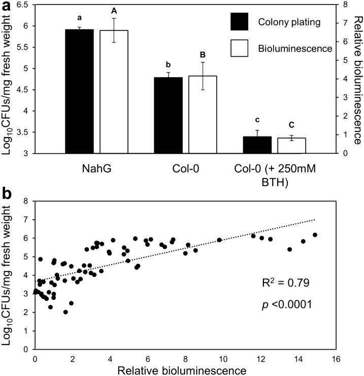
The method presented here uses a bioluminescent Pst DC3000 strain that constitutively expresses the luxCDABE operon from Photorhabdus luminescens (Pst::LUX) and requires a common gel documentation (Gel Doc) system with a sensitive CCD/CMOS camera and imaging software (Photoshop or Image J). By capturing bright field and bioluminescence images from Pst::LUX-infected leaves, we imaged the spatiotemporal dynamics of Pst infection. Analysis of bioluminescence from live Pst bacteria over a 5-day time course after spray inoculation of Arabidopsis revealed transition of the bacterial presence from the older leaves to the younger leaves and apical meristem. Colonisation by Pst:LUX bioluminescence was obtained from digital photos by calculating relative bioluminescence values, which is adjusted for bioluminescence intensity and normalised by leaf surface. This method detected statistically significant differences in Pst::LUX colonisation between Arabidopsis genotypes varying in basal resistance, as well as statistically significant reductions in Pst::LUX colonisation by resistance-inducing treatments in both Arabidopsis and tomato. Comparison of relative bioluminescence values to conventional colony counting on selective agar medium revealed a statistically significant correlation, which was reproducible between different Gel Doc systems.
We present a non-destructive method to quantify colonisation by bioluminescent Pst::LUX in plants. Using a common Gel Doc system and imaging software, our method requires less time and labour than conventional methods that are based on destructive sampling of infected leaf material. Furthermore, in contrast to conventional strategies, our method provides additional information about the spatial-temporal patterns of Pst colonisation.
细菌性叶部病原体丁香假单胞菌番茄致病变种(Pst)是植物病理学研究中最常用的模式病原体。以往研究植物与Pst相互作用的方法依赖于对Pst定殖的破坏性定量分析,这种方法既耗费人力又耗时,且无法对细菌定殖进行时空监测。在此,我们描述了一种快速且非破坏性的方法,用于定量和可视化Pst在拟南芥和番茄完整叶片中的时空定殖情况。
本文介绍的方法使用了一种组成型表达来自发光杆菌属的luxCDABE操纵子的生物发光Pst DC3000菌株(Pst::LUX),并且需要一个配备灵敏的电荷耦合器件/互补金属氧化物半导体(CCD/CMOS)相机和成像软件(Photoshop或Image J)的普通凝胶成像系统。通过拍摄Pst::LUX感染叶片的明场和生物发光图像,我们对Pst感染的时空动态进行了成像。对拟南芥喷雾接种后5天内活的Pst细菌的生物发光分析表明,细菌的存在从老叶转移到了幼叶和顶端分生组织。通过计算相对生物发光值从数码照片中获得Pst:LUX生物发光的定殖情况,该值针对生物发光强度进行了调整,并通过叶面积进行了归一化。该方法检测到基础抗性不同的拟南芥基因型之间Pst::LUX定殖存在统计学上的显著差异,以及在拟南芥和番茄中通过抗性诱导处理导致Pst::LUX定殖有统计学上的显著降低。将相对生物发光值与在选择性琼脂培养基上的传统菌落计数进行比较,发现存在统计学上的显著相关性,并且在不同的凝胶成像系统之间具有可重复性。
我们提出了一种非破坏性方法来定量植物中生物发光Pst::LUX的定殖情况。使用普通的凝胶成像系统和成像软件,我们的方法比基于对感染叶片材料进行破坏性取样的传统方法所需的时间和人力更少。此外,与传统策略不同,我们的方法提供了有关Pst定殖时空模式的额外信息。