• 文献检索
  • 文档翻译
  • 深度研究
  • 学术资讯
  • Suppr Zotero 插件Zotero 插件
  • 邀请有礼
  • 套餐&价格
  • 历史记录
应用&插件
Suppr Zotero 插件Zotero 插件浏览器插件Mac 客户端Windows 客户端微信小程序
定价
高级版会员购买积分包购买API积分包
服务
文献检索文档翻译深度研究API 文档MCP 服务
关于我们
关于 Suppr公司介绍联系我们用户协议隐私条款
关注我们

Suppr 超能文献

核心技术专利:CN118964589B侵权必究
粤ICP备2023148730 号-1Suppr @ 2026

文献检索

告别复杂PubMed语法,用中文像聊天一样搜索,搜遍4000万医学文献。AI智能推荐,让科研检索更轻松。

立即免费搜索

文件翻译

保留排版,准确专业,支持PDF/Word/PPT等文件格式,支持 12+语言互译。

免费翻译文档

深度研究

AI帮你快速写综述,25分钟生成高质量综述,智能提取关键信息,辅助科研写作。

立即免费体验

利用扩散磁共振成像对小鼠大脑进行宏观结构和连接组图谱绘制

Macroscopic Structural and Connectome Mapping of the Mouse Brain Using Diffusion Magnetic Resonance Imaging.

作者信息

Arefin Tanzil Mahmud, Lee Choong Heon, White Jordon D, Zhang Jiangyang, Kaffman Arie

机构信息

Bernard Irene Schwartz Center for Biomedical Imaging, Department of Radiology, New York University Grossman School of Medicine, New York, USA.

Department of Psychiatry, Yale University School of Medicine, New Haven, Connecticut, USA.

出版信息

Bio Protoc. 2021 Nov 20;11(22):e4221. doi: 10.21769/BioProtoc.4221.

DOI:10.21769/BioProtoc.4221
PMID:34909442
原文链接:https://pmc.ncbi.nlm.nih.gov/articles/PMC8635841/
Abstract

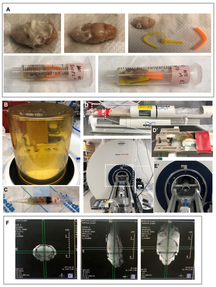
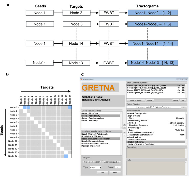
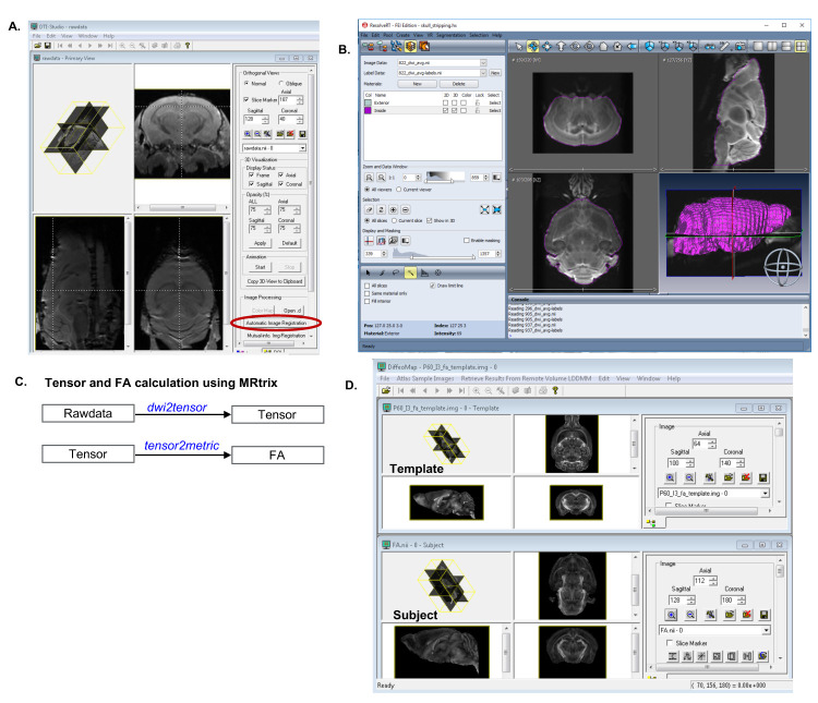
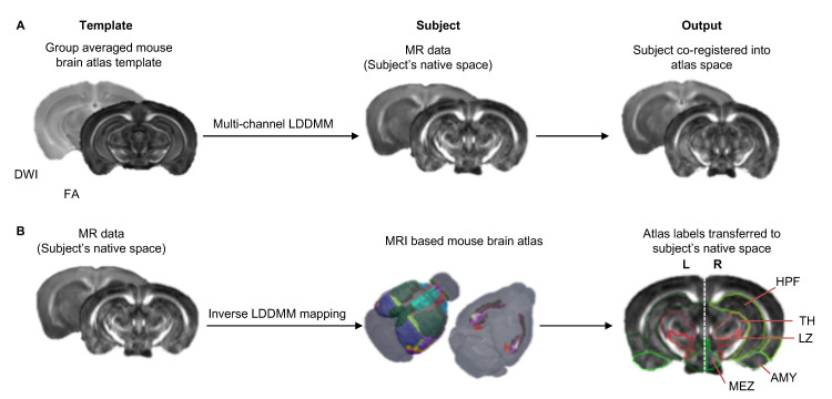
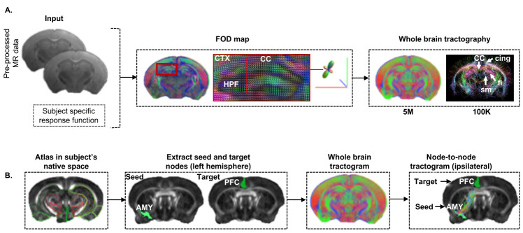
Translational work in rodents elucidates basic mechanisms that drive complex behaviors relevant to psychiatric and neurological conditions. Nonetheless, numerous promising studies in rodents later fail in clinical trials, highlighting the need for improving the translational utility of preclinical studies in rodents. Imaging of small rodents provides an important strategy to address this challenge, as it enables a whole-brain unbiased search for structural and dynamic changes that can be directly compared to human imaging. The functional significance of structural changes identified using imaging can then be further investigated using molecular and genetic tools available for the mouse. Here, we describe a pipeline for unbiased search and characterization of structural changes and network properties, based on diffusion MRI data covering the entire mouse brain at an isotropic resolution of 100 µm. We first used unbiased whole-brain voxel-based analyses to identify volumetric and microstructural alterations in the brain of adult mice exposed to unpredictable postnatal stress (UPS), which is a mouse model of complex early life stress (ELS). Brain regions showing structural abnormalities were used as nodes to generate a grid for assessing structural connectivity and network properties based on graph theory. The technique described here can be broadly applied to understand brain connectivity in other mouse models of human disorders, as well as in genetically modified mouse strains. Graphic abstract: Scale bar = 1 mm.

摘要

在啮齿动物身上开展的转化研究阐明了驱动与精神和神经疾病相关复杂行为的基本机制。尽管如此,许多在啮齿动物身上开展的前景看好的研究后来在临床试验中失败了,这凸显了提高啮齿动物临床前研究转化效用的必要性。对小型啮齿动物进行成像提供了应对这一挑战的重要策略,因为它能够对全脑进行无偏倚搜索,以寻找可直接与人类成像进行比较的结构和动态变化。然后,可以使用小鼠可用的分子和遗传工具进一步研究通过成像确定的结构变化的功能意义。在此,我们描述了一种基于扩散磁共振成像(MRI)数据的流程,用于对结构变化和网络特性进行无偏倚搜索和表征,该数据以100微米的各向同性分辨率覆盖整个小鼠大脑。我们首先使用无偏倚的基于全脑体素的分析方法,来识别暴露于不可预测的产后应激(UPS)的成年小鼠大脑中的体积和微观结构改变,UPS是一种复杂早期生活应激(ELS)的小鼠模型。将显示结构异常的脑区用作节点,以生成一个网格,用于基于图论评估结构连通性和网络特性。这里描述的技术可广泛应用于理解人类疾病的其他小鼠模型以及基因改造小鼠品系中的脑连通性。图形摘要:比例尺 = 1毫米。

https://cdn.ncbi.nlm.nih.gov/pmc/blobs/ffe1/8635841/33979480a1a4/BioProtoc-11-22-4221-g005.jpg
https://cdn.ncbi.nlm.nih.gov/pmc/blobs/ffe1/8635841/808f30382cc8/BioProtoc-11-22-4221-g001.jpg
https://cdn.ncbi.nlm.nih.gov/pmc/blobs/ffe1/8635841/44675c8e6d7f/BioProtoc-11-22-4221-g002.jpg
https://cdn.ncbi.nlm.nih.gov/pmc/blobs/ffe1/8635841/42024073d25f/BioProtoc-11-22-4221-g003.jpg
https://cdn.ncbi.nlm.nih.gov/pmc/blobs/ffe1/8635841/293ec6c6b33c/BioProtoc-11-22-4221-g004.jpg
https://cdn.ncbi.nlm.nih.gov/pmc/blobs/ffe1/8635841/33979480a1a4/BioProtoc-11-22-4221-g005.jpg

相似文献

1
Macroscopic Structural and Connectome Mapping of the Mouse Brain Using Diffusion Magnetic Resonance Imaging.利用扩散磁共振成像对小鼠大脑进行宏观结构和连接组图谱绘制
Bio Protoc. 2021 Nov 20;11(22):e4221. doi: 10.21769/BioProtoc.4221.
2
Large-scale topology and the default mode network in the mouse connectome.小鼠连接组中的大规模拓扑结构与默认模式网络
Proc Natl Acad Sci U S A. 2014 Dec 30;111(52):18745-50. doi: 10.1073/pnas.1404346111. Epub 2014 Dec 15.
3
A Diffusion MRI Tractography Connectome of the Mouse Brain and Comparison with Neuronal Tracer Data.小鼠脑的扩散磁共振成像纤维束连接组及其与神经元示踪剂数据的比较。
Cereb Cortex. 2015 Nov;25(11):4628-37. doi: 10.1093/cercor/bhv121. Epub 2015 Jun 5.
4
Resolution and b value dependent structural connectome in ex vivo mouse brain.离体小鼠脑的分辨率和 b 值相关结构连接组学
Neuroimage. 2022 Jul 15;255:119199. doi: 10.1016/j.neuroimage.2022.119199. Epub 2022 Apr 10.
5
Clinical factors associated with microstructural connectome related brain dysmaturation in term neonates with congenital heart disease.先天性心脏病足月儿中与微观连接组相关脑发育异常相关的临床因素。
Front Neurosci. 2022 Nov 18;16:952355. doi: 10.3389/fnins.2022.952355. eCollection 2022.
6
Mapping Structural Connectivity Using Diffusion MRI: Challenges and Opportunities.利用弥散磁共振成像进行结构连接映射:挑战与机遇。
J Magn Reson Imaging. 2021 Jun;53(6):1666-1682. doi: 10.1002/jmri.27188. Epub 2020 Jun 17.
7
Abnormal structural networks characterize major depressive disorder: a connectome analysis.异常的结构网络是重度抑郁症的特征:连接组学分析。
Biol Psychiatry. 2014 Oct 1;76(7):567-74. doi: 10.1016/j.biopsych.2014.02.018. Epub 2014 Mar 6.
8
The structural-functional connectome and the default mode network of the human brain.人类大脑的结构-功能连接组和默认模式网络。
Neuroimage. 2014 Nov 15;102 Pt 1:142-51. doi: 10.1016/j.neuroimage.2013.09.069. Epub 2013 Oct 4.
9
Connectivity and tissue microstructural alterations in right and left temporal lobe epilepsy revealed by diffusion spectrum imaging.扩散光谱成像揭示的左右颞叶癫痫中的连通性和组织微观结构改变
Neuroimage Clin. 2014 Aug 1;5:349-58. doi: 10.1016/j.nicl.2014.07.013. eCollection 2014.
10
Whole mouse brain structural connectomics using magnetic resonance histology.基于磁共振组织学的全鼠脑结构连接组学
Brain Struct Funct. 2018 Dec;223(9):4323-4335. doi: 10.1007/s00429-018-1750-x. Epub 2018 Sep 17.

引用本文的文献

1
Cerebellar output neurons can impair non-motor behaviors by altering development of extracerebellar connectivity.小脑输出神经元可通过改变小脑外连接的发育来损害非运动行为。
Nat Commun. 2025 Feb 21;16(1):1858. doi: 10.1038/s41467-025-57080-6.
2
Sex-specific signatures of GLP-1 and amylin on resting state brain activity and functional connectivity in awake rats.胰高血糖素样肽-1(GLP-1)和胰淀素对清醒大鼠静息态脑活动及功能连接的性别特异性特征
Neuropharmacology. 2025 May 15;269:110348. doi: 10.1016/j.neuropharm.2025.110348. Epub 2025 Feb 5.
3
Quantifying the Fascicular Changes in Recovered Achilles Tendon Patients Using Diffusion Magnetic Resonance Imaging and Tractography.

本文引用的文献

1
Early life stress causes sex-specific changes in adult fronto-limbic connectivity that differentially drive learning.早期生活压力会导致成年额眶皮质-边缘连接的性别特异性变化,从而不同程度地影响学习能力。
Elife. 2020 Dec 1;9:e58301. doi: 10.7554/eLife.58301.
2
The present and the future of microstructure MRI: From a paradigm shift to normal science.微观结构 MRI 的现状与未来:从范式转变到常规科学。
J Neurosci Methods. 2021 Mar 1;351:108947. doi: 10.1016/j.jneumeth.2020.108947. Epub 2020 Oct 21.
3
Diffusion Tensor Imaging-Based Studies at the Group-Level Applied to Animal Models of Neurodegenerative Diseases.
使用扩散磁共振成像和纤维束成像量化跟腱损伤恢复患者的束状变化。
J Eng Sci Med Diagn Ther. 2025 Aug 1;8(3):031006. doi: 10.1115/1.4066623. Epub 2024 Oct 16.
4
Microglia: The Drunken Gardeners of Early Adversity.小胶质细胞:早期逆境中的醉园丁。
Biomolecules. 2024 Aug 8;14(8):964. doi: 10.3390/biom14080964.
5
Cerebellar output neurons impair non-motor behaviors by altering development of extracerebellar connectivity.小脑输出神经元通过改变小脑外连接的发育来损害非运动行为。
bioRxiv. 2024 Jul 8:2024.07.08.602496. doi: 10.1101/2024.07.08.602496.
6
Early adversity causes sex-specific deficits in perforant pathway connectivity and contextual memory in adolescent mice.早期逆境导致青春期小鼠海马传入通路连接和情景记忆出现性别特异性缺陷。
Biol Sex Differ. 2024 May 7;15(1):39. doi: 10.1186/s13293-024-00616-0.
7
Open-source versatile 3D-print animal conditioning platform design for in vivo preclinical brain imaging in awake mice and anesthetized mice and rats.开源通用 3D 打印动物训练平台设计,用于清醒小鼠和麻醉小鼠及大鼠的活体临床前脑成像。
Lab Anim (NY). 2024 Feb;53(2):33-42. doi: 10.1038/s41684-023-01320-z. Epub 2024 Jan 26.
8
Early adversity changes the economic conditions of mouse structural brain network organization.早期逆境改变了小鼠结构大脑网络组织的经济条件。
Dev Psychobiol. 2023 Sep;65(6):e22405. doi: 10.1002/dev.22405.
9
Towards reliable reconstruction of the mouse brain corticothalamic connectivity using diffusion MRI.使用弥散磁共振成像技术可靠地重建小鼠大脑皮质丘脑连接。
Neuroimage. 2023 Jun;273:120111. doi: 10.1016/j.neuroimage.2023.120111. Epub 2023 Apr 13.
10
Using mesoscopic tract-tracing data to guide the estimation of fiber orientation distributions in the mouse brain from diffusion MRI.利用介观轨迹追踪数据来指导从弥散 MRI 中估计小鼠大脑中的纤维方向分布。
Neuroimage. 2023 Apr 15;270:119999. doi: 10.1016/j.neuroimage.2023.119999. Epub 2023 Mar 5.
基于扩散张量成像的群体水平研究应用于神经退行性疾病动物模型
Front Neurosci. 2020 Aug 31;14:734. doi: 10.3389/fnins.2020.00734. eCollection 2020.
4
Graph theoretical quantification of white matter reorganization after cortical stroke in mice.基于图论的皮质卒中后小鼠脑白质重组的量化研究
Neuroimage. 2020 Aug 15;217:116873. doi: 10.1016/j.neuroimage.2020.116873. Epub 2020 May 5.
5
Altered structural connectivity networks in a mouse model of complete and partial dysgenesis of the corpus callosum.完全和部分胼胝体发育不全小鼠模型中结构连接网络的改变。
Neuroimage. 2020 Aug 15;217:116868. doi: 10.1016/j.neuroimage.2020.116868. Epub 2020 Apr 29.
6
A Student's Guide to Neural Circuit Tracing.神经回路追踪学生指南
Front Neurosci. 2019 Aug 27;13:897. doi: 10.3389/fnins.2019.00897. eCollection 2019.
7
Enhancing the Utility of Preclinical Research in Neuropsychiatry Drug Development.提高临床前研究在神经精神药物研发中的效用。
Methods Mol Biol. 2019;2011:3-22. doi: 10.1007/978-1-4939-9554-7_1.
8
Towards an efficient segmentation of small rodents brain: A short critical review.朝向小型啮齿动物大脑的有效分割:一个简短的批判性回顾。
J Neurosci Methods. 2019 Jul 15;323:82-89. doi: 10.1016/j.jneumeth.2019.05.003. Epub 2019 May 15.
9
Magnetic resonance imaging of mouse brain networks plasticity following motor learning.磁共振成像在运动学习后小鼠脑网络可塑性中的应用。
PLoS One. 2019 May 8;14(5):e0216596. doi: 10.1371/journal.pone.0216596. eCollection 2019.
10
Mapping Histological Slice Sequences to the Allen Mouse Brain Atlas Without 3D Reconstruction.无需三维重建将组织学切片序列映射到艾伦小鼠脑图谱
Front Neuroinform. 2018 Dec 11;12:93. doi: 10.3389/fninf.2018.00093. eCollection 2018.