• 文献检索
  • 文档翻译
  • 深度研究
  • 学术资讯
  • Suppr Zotero 插件Zotero 插件
  • 邀请有礼
  • 套餐&价格
  • 历史记录
应用&插件
Suppr Zotero 插件Zotero 插件浏览器插件Mac 客户端Windows 客户端微信小程序
定价
高级版会员购买积分包购买API积分包
服务
文献检索文档翻译深度研究API 文档MCP 服务
关于我们
关于 Suppr公司介绍联系我们用户协议隐私条款
关注我们

Suppr 超能文献

核心技术专利:CN118964589B侵权必究
粤ICP备2023148730 号-1Suppr @ 2026

文献检索

告别复杂PubMed语法,用中文像聊天一样搜索,搜遍4000万医学文献。AI智能推荐,让科研检索更轻松。

立即免费搜索

文件翻译

保留排版,准确专业,支持PDF/Word/PPT等文件格式,支持 12+语言互译。

免费翻译文档

深度研究

AI帮你快速写综述,25分钟生成高质量综述,智能提取关键信息,辅助科研写作。

立即免费体验

无需三维重建将组织学切片序列映射到艾伦小鼠脑图谱

Mapping Histological Slice Sequences to the Allen Mouse Brain Atlas Without 3D Reconstruction.

作者信息

Xiong Jing, Ren Jing, Luo Liqun, Horowitz Mark

机构信息

Department of Electrical Engineering, Stanford University, Stanford, CA, United States.

Department of Biology, Howard Hughes Medical Institute, Stanford University, Stanford, CA, United States.

出版信息

Front Neuroinform. 2018 Dec 11;12:93. doi: 10.3389/fninf.2018.00093. eCollection 2018.

DOI:10.3389/fninf.2018.00093
PMID:30618698
原文链接:https://pmc.ncbi.nlm.nih.gov/articles/PMC6297281/
Abstract

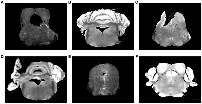
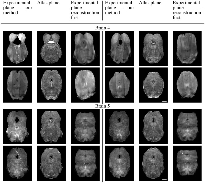
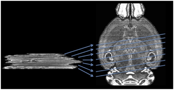
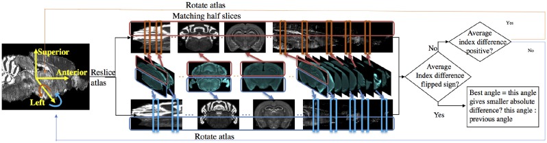
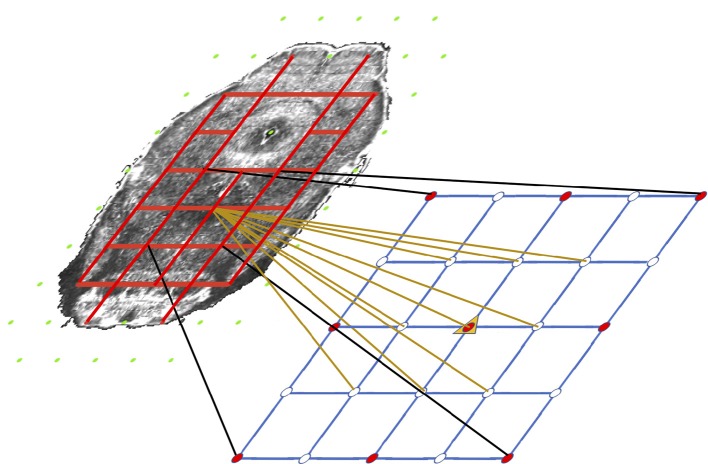
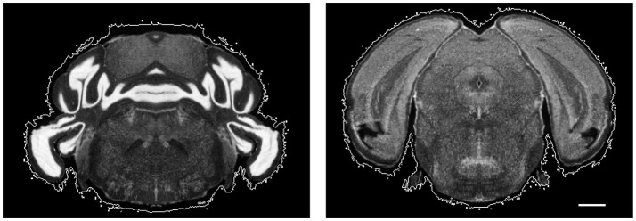
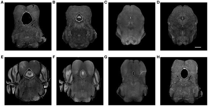
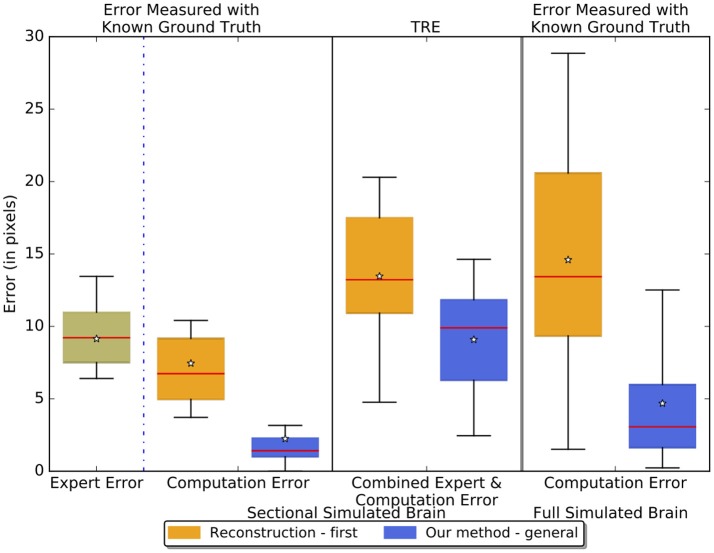
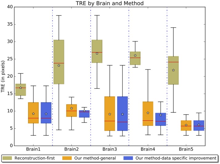
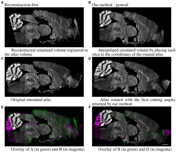
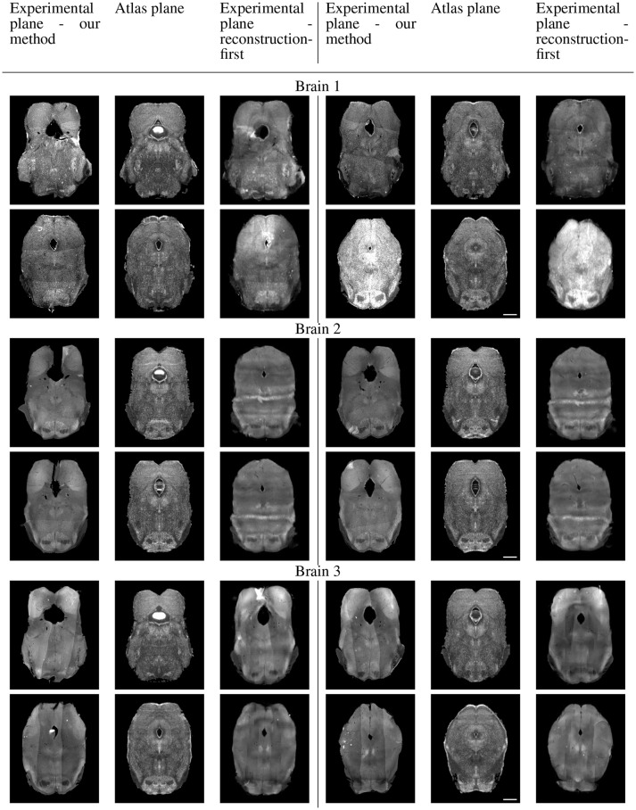
Histological brain slices are widely used in neuroscience to study the anatomical organization of neural circuits. Systematic and accurate comparisons of anatomical data from multiple brains, especially from different studies, can benefit tremendously from registering histological slices onto a common reference atlas. Most existing methods rely on an initial reconstruction of the volume before registering it to a reference atlas. Because these slices are prone to distortions during the sectioning process and often sectioned with non-standard angles, reconstruction is challenging and often inaccurate. Here we describe a framework that maps each slice to its corresponding plane in the Allen Mouse Brain Atlas (2015) to build a plane-wise mapping and then perform 2D nonrigid registration to build a pixel-wise mapping. We use the L2 norm of the histogram of oriented gradients difference of two patches as the similarity metric for both steps and a Markov random field formulation that incorporates tissue coherency to compute the nonrigid registration. To fix significantly distorted regions that are misshaped or much smaller than the control grids, we train a context-aggregation network to segment and warp them to their corresponding regions with thin plate spline. We have shown that our method generates results comparable to an expert neuroscientist and is significantly better than reconstruction-first approaches. Code and sample dataset are available at sites.google.com/view/brain-mapping.

摘要

组织学脑切片在神经科学中被广泛用于研究神经回路的解剖组织。对来自多个大脑(尤其是来自不同研究)的解剖数据进行系统且准确的比较,通过将组织学切片配准到一个通用参考图谱上可极大受益。大多数现有方法在将体积配准到参考图谱之前依赖于其初始重建。由于这些切片在切片过程中容易变形,且常常以非标准角度切片,重建具有挑战性且往往不准确。在此,我们描述了一个框架,该框架将每个切片映射到艾伦小鼠脑图谱(2015 年)中的对应平面以构建平面映射,然后执行二维非刚性配准以构建逐像素映射。我们在两个步骤中均使用两个小块的定向梯度差异直方图的 L2 范数作为相似性度量,并使用结合组织连贯性的马尔可夫随机场公式来计算非刚性配准。为了修复严重扭曲的区域(这些区域形状异常或比控制网格小得多),我们训练了一个上下文聚合网络,用薄板样条将它们分割并扭曲到其对应区域。我们已经表明,我们的方法生成的结果与专业神经科学家的结果相当,并且明显优于先重建的方法。代码和示例数据集可在 sites.google.com/view/brain-mapping 上获取。

https://cdn.ncbi.nlm.nih.gov/pmc/blobs/b8a8/6297281/38de30c3a1cf/fninf-12-00093-g0011.jpg
https://cdn.ncbi.nlm.nih.gov/pmc/blobs/b8a8/6297281/b4ddb9344fa3/fninf-12-00093-g0001.jpg
https://cdn.ncbi.nlm.nih.gov/pmc/blobs/b8a8/6297281/799a1e36aab3/fninf-12-00093-g0002.jpg
https://cdn.ncbi.nlm.nih.gov/pmc/blobs/b8a8/6297281/9083509f393a/fninf-12-00093-g0003.jpg
https://cdn.ncbi.nlm.nih.gov/pmc/blobs/b8a8/6297281/58a5330416e1/fninf-12-00093-g0004.jpg
https://cdn.ncbi.nlm.nih.gov/pmc/blobs/b8a8/6297281/61b4c6cbe807/fninf-12-00093-g0005.jpg
https://cdn.ncbi.nlm.nih.gov/pmc/blobs/b8a8/6297281/9eae4740afca/fninf-12-00093-g0006.jpg
https://cdn.ncbi.nlm.nih.gov/pmc/blobs/b8a8/6297281/bfa1796146e4/fninf-12-00093-g0007.jpg
https://cdn.ncbi.nlm.nih.gov/pmc/blobs/b8a8/6297281/6afe59d3f334/fninf-12-00093-g0008.jpg
https://cdn.ncbi.nlm.nih.gov/pmc/blobs/b8a8/6297281/c6a5d7e31fbb/fninf-12-00093-g0009.jpg
https://cdn.ncbi.nlm.nih.gov/pmc/blobs/b8a8/6297281/ab22e1eecfca/fninf-12-00093-g0010.jpg
https://cdn.ncbi.nlm.nih.gov/pmc/blobs/b8a8/6297281/38de30c3a1cf/fninf-12-00093-g0011.jpg

相似文献

1
Mapping Histological Slice Sequences to the Allen Mouse Brain Atlas Without 3D Reconstruction.无需三维重建将组织学切片序列映射到艾伦小鼠脑图谱
Front Neuroinform. 2018 Dec 11;12:93. doi: 10.3389/fninf.2018.00093. eCollection 2018.
2
Localization and Registration of 2D Histological Mouse Brain Images in 3D Atlas Space.二维组织学小鼠脑图像在三维图谱空间中的定位和配准。
Neuroinformatics. 2023 Jul;21(3):615-630. doi: 10.1007/s12021-023-09632-8. Epub 2023 Jun 26.
3
Robust joint registration of multiple stains and MRI for multimodal 3D histology reconstruction: Application to the Allen human brain atlas.多模态 3D 组织学重建的多染色和 MRI 的稳健联合配准:Allen 人类大脑图谱的应用。
Med Image Anal. 2022 Jan;75:102265. doi: 10.1016/j.media.2021.102265. Epub 2021 Oct 16.
4
Geometry processing of conventionally produced mouse brain slice images.常规制作的小鼠脑切片图像的几何处理。
J Neurosci Methods. 2018 Aug 1;306:45-56. doi: 10.1016/j.jneumeth.2018.04.008. Epub 2018 Apr 22.
5
Slice-to-Volume Nonrigid Registration of Histological Sections to MR Images of the Human Brain.人脑组织学切片到磁共振图像的切片到体积非刚性配准
Anat Res Int. 2011;2011:287860. doi: 10.1155/2011/287860. Epub 2010 Dec 12.
6
giRAff: an automated atlas segmentation tool adapted to single histological slices.giRAff:一种适用于单一组织学切片的自动图谱分割工具。
Front Neurosci. 2024 Jan 11;17:1230814. doi: 10.3389/fnins.2023.1230814. eCollection 2023.
7
Constructing and optimizing 3D atlases from 2D data with application to the developing mouse brain.从二维数据构建和优化 3D 图谱及其在发育中老鼠大脑中的应用。
Elife. 2021 Feb 11;10:e61408. doi: 10.7554/eLife.61408.
8
Precise Mapping of Single Neurons by Calibrated 3D Reconstruction of Brain Slices Reveals Topographic Projection in Mouse Visual Cortex.通过校准的脑片 3D 重建对单个神经元进行精确映射,揭示了小鼠视觉皮层中的拓扑投影。
Cell Rep. 2020 May 26;31(8):107682. doi: 10.1016/j.celrep.2020.107682.
9
The creation of a brain atlas for image guided neurosurgery using serial histological data.利用系列组织学数据创建用于图像引导神经外科手术的脑图谱。
Neuroimage. 2006 Apr 1;30(2):359-76. doi: 10.1016/j.neuroimage.2005.09.041. Epub 2006 Jan 9.
10
Supervised Learning With Perceptual Similarity for Multimodal Gene Expression Registration of a Mouse Brain Atlas.基于感知相似性的监督学习用于小鼠脑图谱的多模态基因表达配准
Front Neuroinform. 2021 Jul 28;15:691918. doi: 10.3389/fninf.2021.691918. eCollection 2021.

引用本文的文献

1
An extended and improved CCFv3 annotation and Nissl atlas of the entire mouse brain.扩展并改进的整个小鼠脑CCFv3注释和尼氏图谱。
Imaging Neurosci (Camb). 2025 May 21;3. doi: 10.1162/imag_a_00565. eCollection 2025.
2
Mapping brain-wide activity networks: brainways as a tool for neurobiological discovery.绘制全脑活动网络:Brainways作为神经生物学发现的工具
Neuropsychopharmacology. 2025 Apr 22. doi: 10.1038/s41386-025-02105-3.
3
Multimodal Mass Spectrometry Imaging in Atlas Building: A Review.图谱构建中的多模态质谱成像:综述

本文引用的文献

1
A Survey of Methods for 3D Histology Reconstruction.三维组织学重建方法研究综述。
Med Image Anal. 2018 May;46:73-105. doi: 10.1016/j.media.2018.02.004. Epub 2018 Feb 21.
2
3D volume reconstruction from serial breast specimen radiographs for mapping between histology and 3D whole specimen imaging.通过乳腺标本连续X光片进行3D体积重建,用于组织学与3D全标本成像之间的映射。
Med Phys. 2017 Mar;44(3):935-948. doi: 10.1002/mp.12077.
3
NBLAST: Rapid, Sensitive Comparison of Neuronal Structure and Construction of Neuron Family Databases.
Semin Nephrol. 2024 Nov;44(6):151578. doi: 10.1016/j.semnephrol.2025.151578. Epub 2025 Apr 16.
4
Bell Jar: A Semiautomated Registration and Cell Counting Tool for Mouse Neurohistology Analysis.钟形罩:一种用于小鼠神经组织学分析的半自动配准和细胞计数工具。
eNeuro. 2025 Feb 5;12(2). doi: 10.1523/ENEURO.0036-23.2025. Print 2025 Feb.
5
giRAff: an automated atlas segmentation tool adapted to single histological slices.giRAff:一种适用于单一组织学切片的自动图谱分割工具。
Front Neurosci. 2024 Jan 11;17:1230814. doi: 10.3389/fnins.2023.1230814. eCollection 2023.
6
D-LMBmap: a fully automated deep-learning pipeline for whole-brain profiling of neural circuitry.D-LMBmap:一个用于全脑神经回路分析的全自动深度学习管道。
Nat Methods. 2023 Oct;20(10):1593-1604. doi: 10.1038/s41592-023-01998-6. Epub 2023 Sep 28.
7
DeepSlice: rapid fully automatic registration of mouse brain imaging to a volumetric atlas.DeepSlice:快速全自动将小鼠脑成像与容积图谱进行配准。
Nat Commun. 2023 Sep 21;14(1):5884. doi: 10.1038/s41467-023-41645-4.
8
Localization and Registration of 2D Histological Mouse Brain Images in 3D Atlas Space.二维组织学小鼠脑图像在三维图谱空间中的定位和配准。
Neuroinformatics. 2023 Jul;21(3):615-630. doi: 10.1007/s12021-023-09632-8. Epub 2023 Jun 26.
9
SMDB: a Spatial Multimodal Data Browser.SMDB:一个空间多模态数据浏览器。
Nucleic Acids Res. 2023 Jul 5;51(W1):W553-W559. doi: 10.1093/nar/gkad413.
10
Network Neuroscience Untethered: Brain-Wide Immediate Early Gene Expression for the Analysis of Functional Connectivity in Freely Behaving Animals.不受束缚的网络神经科学:用于分析自由行为动物功能连接性的全脑即时早期基因表达
Biology (Basel). 2022 Dec 24;12(1):34. doi: 10.3390/biology12010034.
NBLAST:神经元结构的快速、灵敏比较及神经元家族数据库构建
Neuron. 2016 Jul 20;91(2):293-311. doi: 10.1016/j.neuron.2016.06.012. Epub 2016 Jun 30.
4
Brain-Wide Mapping of Axonal Connections: Workflow for Automated Detection and Spatial Analysis of Labeling in Microscopic Sections.轴突连接的全脑图谱:显微镜切片中标记的自动检测和空间分析工作流程
Front Neuroinform. 2016 Apr 19;10:11. doi: 10.3389/fninf.2016.00011. eCollection 2016.
5
Possum-A Framework for Three-Dimensional Reconstruction of Brain Images from Serial Sections.Possum:一种用于从连续切片进行脑图像三维重建的框架。
Neuroinformatics. 2016 Jul;14(3):265-78. doi: 10.1007/s12021-015-9286-1.
6
Morphological maturation of the mouse brain: An in vivo MRI and histology investigation.小鼠大脑形态成熟度的研究:体内 MRI 和组织学研究。
Neuroimage. 2016 Jan 15;125:144-152. doi: 10.1016/j.neuroimage.2015.10.009. Epub 2015 Oct 14.
7
Feeding the human brain model.为人类大脑模型提供输入。
Curr Opin Neurobiol. 2015 Jun;32:107-14. doi: 10.1016/j.conb.2015.02.003. Epub 2015 Feb 26.
8
The Scalable Brain Atlas: Instant Web-Based Access to Public Brain Atlases and Related Content.可扩展脑图谱:基于网络即时访问公共脑图谱及相关内容。
Neuroinformatics. 2015 Jul;13(3):353-66. doi: 10.1007/s12021-014-9258-x.
9
Automated anatomical interpretation of ion distributions in tissue: linking imaging mass spectrometry to curated atlases.组织中离子分布的自动解剖学解读:将成像质谱与精选图谱相联系。
Anal Chem. 2014 Sep 16;86(18):8974-82. doi: 10.1021/ac502838t. Epub 2014 Aug 25.
10
Automatic generic registration of mass spectrometry imaging data to histology using nonlinear stochastic embedding.基于非线性随机嵌入的质谱成像数据自动泛型配准到组织学。
Anal Chem. 2014 Sep 16;86(18):9204-11. doi: 10.1021/ac502170f. Epub 2014 Aug 26.