Center for In Vivo Microscopy, Department of Radiology, Duke University Medical Center, Durham, NC, United States of America.
Department of Neurology, Duke University Medical Center, Durham, NC, United States of America.
PLoS One. 2019 May 8;14(5):e0216596. doi: 10.1371/journal.pone.0216596. eCollection 2019.
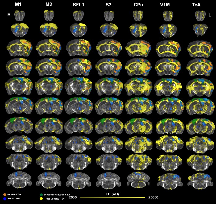
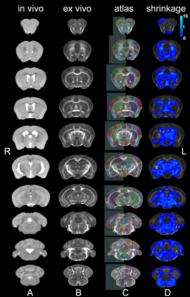
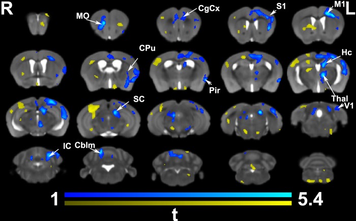
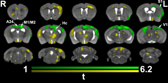
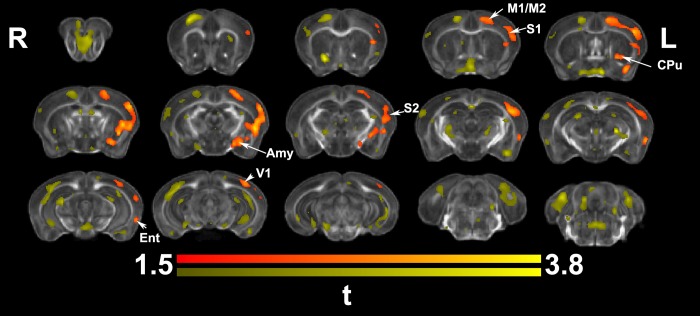
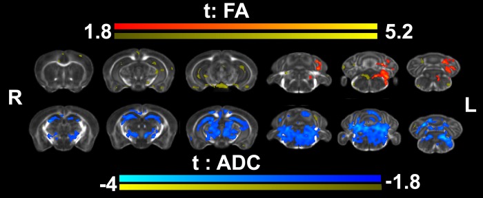
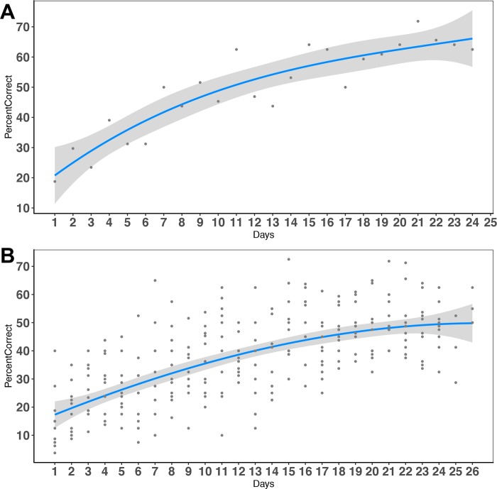
We do not have a full understanding of the mechanisms underlying plasticity in the human brain. Mouse models have well controlled environments and genetics, and provide tools to help dissect the mechanisms underlying the observed responses to therapies devised for humans recovering from injury of ischemic nature or trauma. We aimed to detect plasticity following learning of a unilateral reaching movement, and relied on MRI performed with a rapid structural protocol suitable for in vivo brain imaging, and a longer diffusion tensor imaging (DTI) protocol executed ex vivo. In vivo MRI detected contralateral volume increases in trained animals (reachers), in circuits involved in motor control, sensory processing, and importantly, learning and memory. The temporal association area, parafascicular and mediodorsal thalamic nuclei were also enlarged. In vivo MRI allowed us to detect longitudinal effects over the ~25 days training period. The interaction between time and group (trained versus not trained) supported a role for the contralateral, but also the ipsilateral hemisphere. While ex vivo imaging was affected by shrinkage due to the fixation, it allowed for superior resolution and improved contrast to noise ratios, especially for subcortical structures. We examined microstructural changes based on DTI, and identified increased fractional anisotropy and decreased apparent diffusion coefficient, predominantly in the cerebellum and its connections. Cortical thickness differences did not survive multiple corrections, but uncorrected statistics supported the contralateral effects seen with voxel based volumetric analysis, showing thickening in the somatosensory, motor and visual cortices. In vivo and ex vivo analyses identified plasticity in circuits relevant to selecting actions in a sensory-motor context, through exploitation of learned association and decision making. By mapping a connectivity atlas into our ex vivo template we revealed that changes due to skilled motor learning occurred in a network of 35 regions, including the primary and secondary motor (M1, M2) and sensory cortices (S1, S2), the caudate putamen (CPu), visual (V1) and temporal association cortex. The significant clusters intersected tractography based networks seeded in M1, M2, S1, V1 and CPu at levels > 80%. We found that 89% of the significant cluster belonged to a network seeded in the contralateral M1, and 85% to one seeded in the contralateral M2. Moreover, 40% of the M1 and S1 cluster by network intersections were in the top 80th percentile of the tract densities for their respective networks. Our investigation may be relevant to studies of rehabilitation and recovery, and points to widespread network changes that accompany motor learning that may have potential applications to designing recovery strategies following brain injury.
我们尚未完全了解人类大脑可塑性的形成机制。老鼠模型具有可控的环境和遗传条件,为我们提供了工具,帮助我们剖析人类大脑对各种疗法的反应机制,这些疗法旨在治疗因缺血性损伤或创伤而康复的患者。我们的目的是检测学习单侧伸展运动后的可塑性,我们依赖于使用快速结构协议进行的 MRI 成像,该协议适用于体内大脑成像,以及更长的扩散张量成像(DTI)协议,该协议在体外用。体内 MRI 检测到受过训练的动物(伸展者)中与运动控制、感觉处理以及重要的学习和记忆相关的回路的对侧体积增加。临时关联区域、旁束和内侧背侧丘脑核也增大了。体内 MRI 使我们能够在大约 25 天的训练期间检测到纵向影响。时间与组(训练与未训练)之间的相互作用支持了对侧半球,也支持了同侧半球的作用。虽然体外成像受到固定导致的收缩的影响,但它可以提供更高的分辨率并提高对比噪声比,尤其是对于皮质下结构。我们基于 DTI 检查了微观结构的变化,并确定了分数各向异性的增加和表观扩散系数的降低,主要在小脑及其连接中。皮质厚度差异未通过多次校正而保留,但未经校正的统计数据支持了基于体素体积分析所见的对侧影响,显示出感觉运动皮层的增厚。体内和体外分析均通过利用学习关联和决策制定来鉴定与在感觉运动环境中选择动作相关的回路的可塑性。通过将连接图谱映射到我们的体外模板中,我们揭示了由于熟练的运动学习而导致的变化发生在一个由 35 个区域组成的网络中,包括初级和次级运动(M1、M2)和感觉皮层(S1、S2)、尾状核(CPu)、视觉(V1)和颞部关联皮层。显著的簇与基于 M1、M2、S1、V1 和 CPu 的种子的纤维束追踪网络相交,水平>80%。我们发现,89%的显著簇属于以对侧 M1 为种子的网络,85%的簇属于以对侧 M2 为种子的网络。此外,网络交叉的 M1 和 S1 簇的 40%处于各自网络的束密度的前 80%。我们的研究可能与康复和恢复的研究有关,并指出伴随运动学习的广泛网络变化,这些变化可能对设计脑损伤后的恢复策略具有潜在应用。