Cancer Physiology Department, H. Lee Moffitt Cancer Center and Research Institute, 12902 USF Magnolia Dr, Tampa, FL, 33612, USA.
Translation Imaging Department, Merck & Co, West Point, PA, USA.
Radiat Oncol. 2021 Dec 15;16(1):237. doi: 10.1186/s13014-021-01957-5.
Magnetic Resonance Image guided Stereotactic body radiotherapy (MRgRT) is an emerging technology that is increasingly used in treatment of visceral cancers, such as pancreatic adenocarcinoma (PDAC). Given the variable response rates and short progression times of PDAC, there is an unmet clinical need for a method to assess early RT response that may allow better prescription personalization. We hypothesize that quantitative image feature analysis (radiomics) of the longitudinal MR scans acquired before and during MRgRT may be used to extract information related to early treatment response.
Histogram and texture radiomic features (n = 73) were extracted from the Gross Tumor Volume (GTV) in 0.35T MRgRT scans of 26 locally advanced and borderline resectable PDAC patients treated with 50 Gy RT in 5 fractions. Feature ratios between first (F1) and last (F5) fraction scan were correlated with progression free survival (PFS). Feature stability was assessed through region of interest (ROI) perturbation.
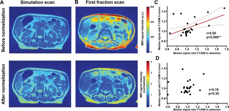
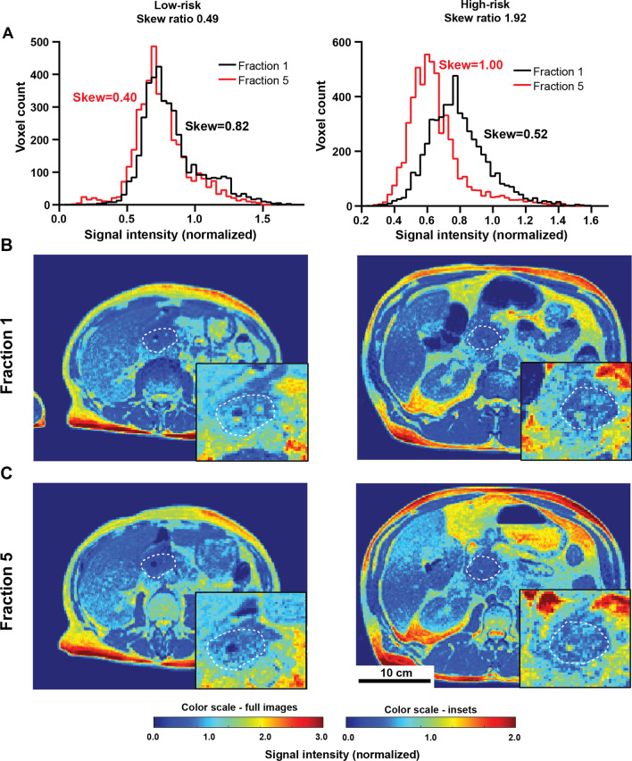
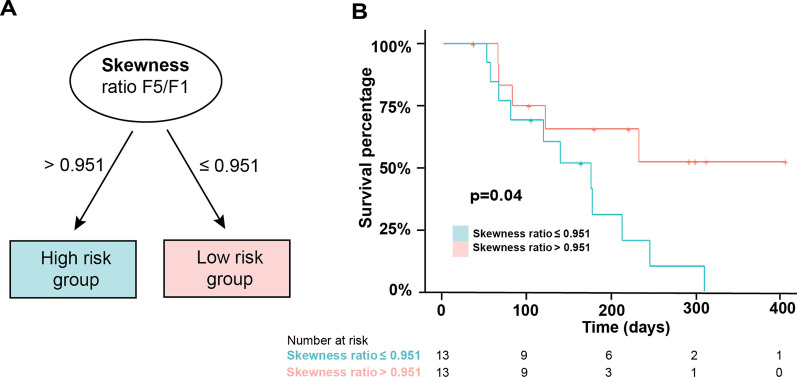
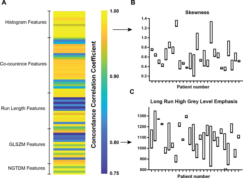
Linear normalization of image intensity to median kidney value showed improved reproducibility of feature quantification. Histogram skewness change during treatment showed significant association with PFS (p = 0.005, HR = 2.75), offering a potential predictive biomarker of RT response. Stability analyses revealed a wide distribution of feature sensitivities to ROI delineation and was able to identify features that were robust to variability in contouring.
This study presents a proof-of-concept for the use of quantitative image analysis in MRgRT for treatment response prediction and providing an analysis pipeline that can be utilized in future MRgRT radiomic studies.
磁共振图像引导立体定向体放射治疗(MRgRT)是一种新兴技术,越来越多地用于治疗内脏癌,如胰腺腺癌(PDAC)。鉴于 PDAC 的反应率和进展时间各不相同,因此需要一种评估早期 RT 反应的方法,以实现更好的处方个性化。我们假设,在接受 MRgRT 之前和期间获取的纵向磁共振扫描的定量图像特征分析(放射组学)可用于提取与早期治疗反应相关的信息。
从 26 例局部晚期和边界可切除 PDAC 患者的 50 Gy 5 次分割 RT 治疗中提取的 0.35T MRgRT 扫描的 Gross Tumor Volume(GTV)中提取了直方图和纹理放射组学特征(n=73)。首次(F1)和最后一次(F5)分次扫描之间的特征比值与无进展生存期(PFS)相关。通过感兴趣区域(ROI)扰动评估特征稳定性。
图像强度的线性归一化到中位数肾值提高了特征量化的可重复性。治疗过程中直方图偏度的变化与 PFS 有显著相关性(p=0.005,HR=2.75),这为 RT 反应提供了一个潜在的预测生物标志物。稳定性分析显示,特征对 ROI 勾画的变化具有广泛的敏感性分布,并且能够识别对勾画变化具有鲁棒性的特征。
本研究提出了在 MRgRT 中使用定量图像分析进行治疗反应预测的概念验证,并提供了一种可用于未来 MRgRT 放射组学研究的分析流程。