• 文献检索
  • 文档翻译
  • 深度研究
  • 学术资讯
  • Suppr Zotero 插件Zotero 插件
  • 邀请有礼
  • 套餐&价格
  • 历史记录
应用&插件
Suppr Zotero 插件Zotero 插件浏览器插件Mac 客户端Windows 客户端微信小程序
定价
高级版会员购买积分包购买API积分包
服务
文献检索文档翻译深度研究API 文档MCP 服务
关于我们
关于 Suppr公司介绍联系我们用户协议隐私条款
关注我们

Suppr 超能文献

核心技术专利:CN118964589B侵权必究
粤ICP备2023148730 号-1Suppr @ 2026

文献检索

告别复杂PubMed语法,用中文像聊天一样搜索,搜遍4000万医学文献。AI智能推荐,让科研检索更轻松。

立即免费搜索

文件翻译

保留排版,准确专业,支持PDF/Word/PPT等文件格式,支持 12+语言互译。

免费翻译文档

深度研究

AI帮你快速写综述,25分钟生成高质量综述,智能提取关键信息,辅助科研写作。

立即免费体验

利用光学相干断层扫描技术评估脉络膜厚度:综述

Evaluation of Choroidal Thickness Using Optical Coherent Tomography: A Review.

作者信息

Xie Rui, Qiu Bingjie, Chhablani Jay, Zhang Xinyuan

机构信息

Beijing Institute of Ophthalmology, Beijing Tongren Eye Center, Tongren Hospital, Capital Medical University, Beijing, China.

Beijing Retinal and Choroidal Vascular Diseases Study Group, Beijing, China.

出版信息

Front Med (Lausanne). 2021 Dec 3;8:783519. doi: 10.3389/fmed.2021.783519. eCollection 2021.

DOI:10.3389/fmed.2021.783519
PMID:34926529
原文链接:https://pmc.ncbi.nlm.nih.gov/articles/PMC8677938/
Abstract

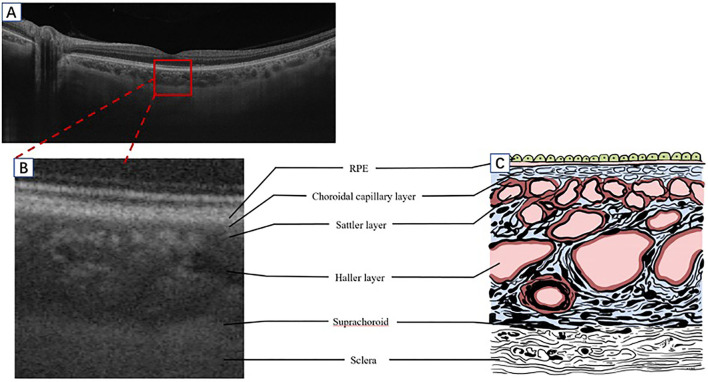
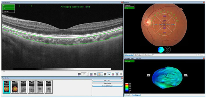
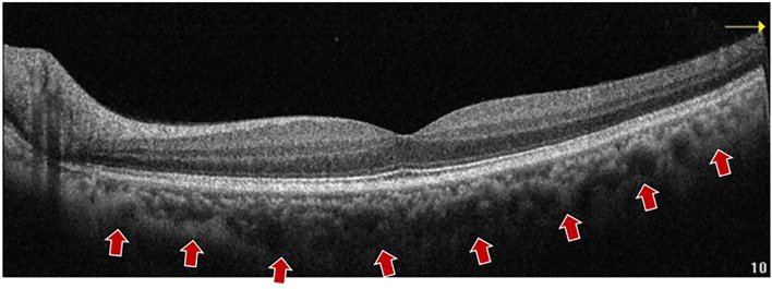
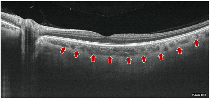
The choroid is the main source of blood and nourishment supply to the eye. The dysfunction of the choroid has been implicated in various retinal and choroidal diseases. The identification and in-depth understanding of pachychoroid spectrum disorders are based on the tremendous progress of optical coherence tomography (OCT) technology in recent years, although visibility of choroid is challenging in the era of the time or spectral domain OCT. The recent rapid revolution of OCTs, such as the enhanced depth imaging OCT and the swept-source OCT, has greatly contributed to the significant improvement in the analysis of the morphology and physiology of the choroid precisely, especially to the choroid-scleral boundary and vasculature. The present review highlights the recently available evidence on the measurement methodology and the clinical significance of choroidal thickness in retinal or choroidal disorders.

摘要

脉络膜是眼球血液和营养供应的主要来源。脉络膜功能障碍与多种视网膜和脉络膜疾病有关。尽管在时域或频域光学相干断层扫描(OCT)时代,脉络膜的可视化具有挑战性,但近年来光学相干断层扫描(OCT)技术的巨大进步为厚脉络膜谱系疾病的识别和深入理解奠定了基础。OCT的最新快速发展,如增强深度成像OCT和扫频源OCT,极大地促进了对脉络膜形态和生理学分析的显著改善,特别是对脉络膜-巩膜边界和脉管系统的分析。本综述重点介绍了目前关于视网膜或脉络膜疾病中脉络膜厚度测量方法和临床意义的现有证据。

https://cdn.ncbi.nlm.nih.gov/pmc/blobs/8b6d/8677938/817603f47e2b/fmed-08-783519-g0005.jpg
https://cdn.ncbi.nlm.nih.gov/pmc/blobs/8b6d/8677938/2afc114dcd37/fmed-08-783519-g0001.jpg
https://cdn.ncbi.nlm.nih.gov/pmc/blobs/8b6d/8677938/cb01c270b9ba/fmed-08-783519-g0002.jpg
https://cdn.ncbi.nlm.nih.gov/pmc/blobs/8b6d/8677938/112465ee7687/fmed-08-783519-g0003.jpg
https://cdn.ncbi.nlm.nih.gov/pmc/blobs/8b6d/8677938/c61ed8070f64/fmed-08-783519-g0004.jpg
https://cdn.ncbi.nlm.nih.gov/pmc/blobs/8b6d/8677938/817603f47e2b/fmed-08-783519-g0005.jpg

相似文献

1
Evaluation of Choroidal Thickness Using Optical Coherent Tomography: A Review.利用光学相干断层扫描技术评估脉络膜厚度:综述
Front Med (Lausanne). 2021 Dec 3;8:783519. doi: 10.3389/fmed.2021.783519. eCollection 2021.
2
EN FACE IMAGING OF PACHYCHOROID SPECTRUM DISORDERS WITH SWEPT-SOURCE OPTICAL COHERENCE TOMOGRAPHY.使用扫频光学相干断层扫描对厚脉络膜疾病谱进行正面成像
Retina. 2016 Mar;36(3):499-516. doi: 10.1097/IAE.0000000000000742.
3
Three-dimensional segmentation and depth-encoded visualization of choroidal vasculature using swept-source optical coherence tomography.应用扫频源光学相干断层扫描对脉络膜血管进行三维分割和深度编码可视化。
Exp Biol Med (Maywood). 2021 Oct;246(20):2238-2245. doi: 10.1177/15353702211028540. Epub 2021 Jul 14.
4
Choroidal analysis in healthy eyes using swept-source optical coherence tomography compared to spectral domain optical coherence tomography.应用扫频源光学相干断层扫描与频域光学相干断层扫描对健康眼的脉络膜进行分析。
Am J Ophthalmol. 2014 Jun;157(6):1272-1281.e1. doi: 10.1016/j.ajo.2014.02.034. Epub 2014 Feb 18.
5
Past, Present, and Future Concepts of the Choroidal Scleral Interface Morphology on Optical Coherence Tomography.光学相干断层扫描技术下脉络膜巩膜界面形态的过去、现在和未来概念
Asia Pac J Ophthalmol (Phila). 2017 Jan-Feb;6(1):94-103. doi: 10.22608/APO.201698.
6
Investigating the choriocapillaris and choroidal vasculature with new optical coherence tomography technologies.利用新型光学相干断层扫描技术研究脉络膜毛细血管和脉络膜血管。
Prog Retin Eye Res. 2016 May;52:130-55. doi: 10.1016/j.preteyeres.2015.10.002. Epub 2015 Oct 23.
7
Optical Coherence Tomography in Inflammatory and Neoplastic Lesions Deforming the Choroidal Profile.光学相干断层扫描在使脉络膜轮廓变形的炎症性和肿瘤性病变中的应用
Diagnostics (Basel). 2023 Jun 7;13(12):1991. doi: 10.3390/diagnostics13121991.
8
Comparison of choroidal thickness measurements between spectral domain optical coherence tomography and swept source optical coherence tomography in children.比较儿童脉络膜厚度测量在光谱域光学相干断层扫描和扫频源光学相干断层扫描之间的差异。
Sci Rep. 2021 Jul 2;11(1):13749. doi: 10.1038/s41598-021-92980-9.
9
Imaging-based Assessment of Choriocapillaris: A Comprehensive Review.基于影像的脉络膜毛细血管评估:全面综述。
Semin Ophthalmol. 2023 Jul;38(5):405-426. doi: 10.1080/08820538.2022.2109939. Epub 2022 Aug 18.
10
Relationship of ocular and systemic factors to the visibility of choroidal-scleral interface using spectral domain optical coherence tomography.使用光谱域光学相干断层扫描技术分析眼部及全身因素与脉络膜-巩膜界面可视性的关系。
Acta Ophthalmol. 2016 Mar;94(2):e142-9. doi: 10.1111/aos.12777. Epub 2015 Jun 24.

引用本文的文献

1
Axicons for improved resolution and depth of focus in adaptive optics scanning light ophthalmoscopy.用于自适应光学扫描激光检眼镜中提高分辨率和焦深的轴棱锥。
Biomed Opt Express. 2025 Jul 16;16(8):3172-3193. doi: 10.1364/BOE.564398. eCollection 2025 Aug 1.
2
Relationship of Choroidal Thickness With Age-Related Macular Degeneration in Asians: An Asian Eye Epidemiology Consortium Meta-Analysis.亚洲人群脉络膜厚度与年龄相关性黄斑变性的关系:亚洲眼流行病学联盟荟萃分析
Invest Ophthalmol Vis Sci. 2025 Jun 2;66(6):36. doi: 10.1167/iovs.66.6.36.
3
Three-Dimensional Choroidal Contour Mapping in Healthy and Diseased Eyes.

本文引用的文献

1
A case of bilateral pachychoroid disease: polypoidal choroidal vasculopathy in one eye and peripheral exudative hemorrhagic chorioretinopathy in contralateral eye.双眼厚脉络膜病变 1 例:一眼为息肉样脉络膜血管病变,对侧眼为周边渗出性出血性脉络膜视网膜病变。
BMC Ophthalmol. 2021 Sep 4;21(1):320. doi: 10.1186/s12886-021-02067-2.
2
Retinal and Choroidal Thickness in relation to C-Reactive Protein on Swept-Source Optical Coherence Tomography.基于扫频源光相干断层扫描的 C 反应蛋白与视网膜和脉络膜厚度的关系。
J Immunol Res. 2021 Jan 25;2021:6628224. doi: 10.1155/2021/6628224. eCollection 2021.
3
Morphologic Features of Myopic Choroidal Neovascularization in Pathologic Myopia on Swept-Source Optical Coherence Tomography.
健康与患病眼睛的三维脉络膜轮廓测绘
Transl Vis Sci Technol. 2025 Jun 2;14(6):16. doi: 10.1167/tvst.14.6.16.
4
Characteristics of the retinal and choroidal thicknesses in myopic young adult males using swept-source optical coherence tomography.使用扫频源光学相干断层扫描技术对近视年轻成年男性视网膜和脉络膜厚度特征的研究
PeerJ. 2025 Mar 5;13:e19030. doi: 10.7717/peerj.19030. eCollection 2025.
5
Ultra-Wide-Field OCT Measurements in Patients with Age-Related Macular Degeneration in Relation to Their Visual Function.年龄相关性黄斑变性患者超广角光学相干断层扫描测量与其视觉功能的关系
Diagnostics (Basel). 2024 Dec 20;14(24):2868. doi: 10.3390/diagnostics14242868.
6
Assessment of choroidal vessels in healthy eyes using 3-dimensional vascular maps and a semi-automated deep learning approach.使用三维血管图和半自动深度学习方法评估健康眼睛中的脉络膜血管。
Sci Rep. 2025 Jan 3;15(1):714. doi: 10.1038/s41598-025-85189-7.
7
Associations of Retinal Vessel Geometry and Optical Coherence Tomography Angiography Metrics With Choroidal Metrics in Diabetic Retinopathy.糖尿病视网膜病变中视网膜血管几何形状和光相干断层扫描血管造影测量值与脉络膜测量值的相关性。
Invest Ophthalmol Vis Sci. 2024 Nov 4;65(13):31. doi: 10.1167/iovs.65.13.31.
8
Choroidal Features of Healthy Iranian Individuals.健康伊朗人的脉络膜特征。
J Curr Ophthalmol. 2024 Aug 10;35(4):369-374. doi: 10.4103/joco.joco_116_23. eCollection 2023 Oct-Dec.
9
Coefficient of spatial variance of choroidal thickness on swept-source optical coherence tomography in healthy eyes.健康眼脉络膜厚度的扫频源光相干断层扫描的空间变异性系数。
Int Ophthalmol. 2024 Jul 8;44(1):318. doi: 10.1007/s10792-024-03218-2.
10
Comparison of manual and artificial intelligence-automated choroidal thickness segmentation of optical coherence tomography imaging in myopic adults.近视成年人中光学相干断层扫描成像的手动与人工智能自动脉络膜厚度分割比较
Eye Vis (Lond). 2024 Jun 3;11(1):21. doi: 10.1186/s40662-024-00385-2.
扫频源光学相干断层扫描下病理性近视中近视性脉络膜新生血管的形态学特征
Front Med (Lausanne). 2020 Dec 23;7:615902. doi: 10.3389/fmed.2020.615902. eCollection 2020.
4
PERIPHERAL EXUDATIVE HEMORRHAGIC CHORIORETINOPATHY-A NEW ADDITION TO THE SPECTRUM OF PACHYCHOROID DISEASE?周边渗出性出血性脉络膜视网膜病变——厚脉络膜疾病谱中的新成员?
Retina. 2021 Jul 1;41(7):1518-1525. doi: 10.1097/IAE.0000000000003063.
5
Pachychoroid spectrum disease.厚脉络膜谱系疾病
Acta Ophthalmol. 2021 Sep;99(6):e806-e822. doi: 10.1111/aos.14683. Epub 2020 Nov 30.
6
Ocular Vascular Changes in Mild Alzheimer's Disease Patients: Foveal Avascular Zone, Choroidal Thickness, and ONH Hemoglobin Analysis.轻度阿尔茨海默病患者的眼部血管变化:中心凹无血管区、脉络膜厚度及视盘血红蛋白分析
J Pers Med. 2020 Nov 15;10(4):231. doi: 10.3390/jpm10040231.
7
Choroidal imaging in patients with Cushing syndrome.库欣综合征患者的脉络膜成像。
Acta Ophthalmol. 2021 Aug;99(5):533-537. doi: 10.1111/aos.14664. Epub 2020 Nov 16.
8
The Eye as a Non-Invasive Window to the Microcirculation in Liver Cirrhosis: A Prospective Pilot Study.眼睛作为肝硬化微循环的无创窗口:一项前瞻性试点研究。
J Clin Med. 2020 Oct 17;9(10):3332. doi: 10.3390/jcm9103332.
9
Pachychoroid: current concepts on clinical features and pathogenesis.厚脉络膜病变:关于临床特征和发病机制的当前概念
Graefes Arch Clin Exp Ophthalmol. 2021 Jun;259(6):1385-1400. doi: 10.1007/s00417-020-04940-0. Epub 2020 Oct 15.
10
Choroidal thickness predicts progression of myopic maculopathy in high myopes: a 2-year longitudinal study.脉络膜厚度预测高度近视性黄斑病变的进展:一项为期 2 年的纵向研究。
Br J Ophthalmol. 2021 Dec;105(12):1744-1750. doi: 10.1136/bjophthalmol-2020-316866. Epub 2020 Sep 24.