• 文献检索
  • 文档翻译
  • 深度研究
  • 学术资讯
  • Suppr Zotero 插件Zotero 插件
  • 邀请有礼
  • 套餐&价格
  • 历史记录
应用&插件
Suppr Zotero 插件Zotero 插件浏览器插件Mac 客户端Windows 客户端微信小程序
定价
高级版会员购买积分包购买API积分包
服务
文献检索文档翻译深度研究API 文档MCP 服务
关于我们
关于 Suppr公司介绍联系我们用户协议隐私条款
关注我们

Suppr 超能文献

核心技术专利:CN118964589B侵权必究
粤ICP备2023148730 号-1Suppr @ 2026

文献检索

告别复杂PubMed语法,用中文像聊天一样搜索,搜遍4000万医学文献。AI智能推荐,让科研检索更轻松。

立即免费搜索

文件翻译

保留排版,准确专业,支持PDF/Word/PPT等文件格式,支持 12+语言互译。

免费翻译文档

深度研究

AI帮你快速写综述,25分钟生成高质量综述,智能提取关键信息,辅助科研写作。

立即免费体验

早期小胶质细胞变化与链脲佐菌素诱导糖尿病大鼠糖尿病视网膜病变的关系。

Early Microglial Changes Associated with Diabetic Retinopathy in Rats with Streptozotocin-Induced Diabetes.

机构信息

Department of Ophthalmology and Visual Science, Seoul St. Mary's Hospital, College of Medicine, The Catholic University of Korea, Seoul, Republic of Korea.

Catholic Institute for Visual Science, College of Medicine, The Catholic University of Korea, Seoul, Republic of Korea.

出版信息

J Diabetes Res. 2021 Dec 8;2021:4920937. doi: 10.1155/2021/4920937. eCollection 2021.

DOI:10.1155/2021/4920937
PMID:34926698
原文链接:https://pmc.ncbi.nlm.nih.gov/articles/PMC8674052/
Abstract

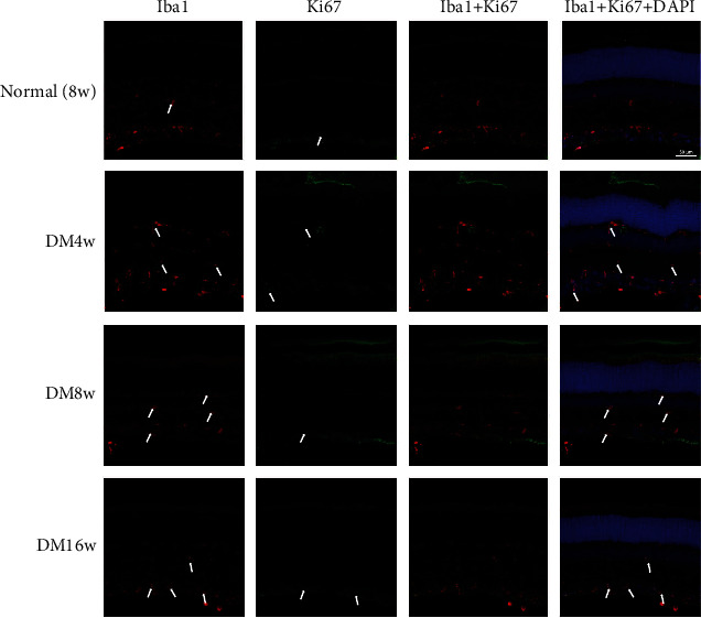
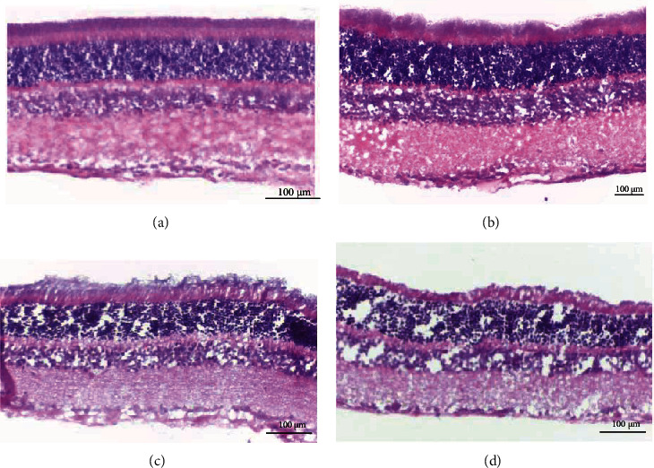
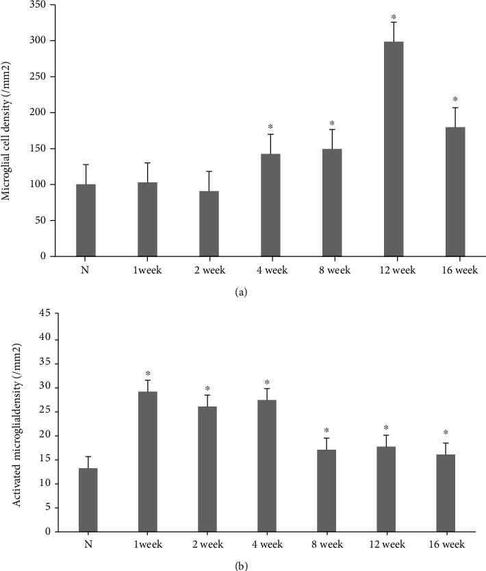
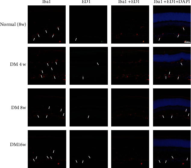
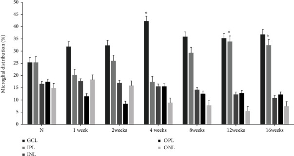
Although morphological changes in microglia have been reported to be associated with diabetic retinopathy, little is known about the early changes in the microglia and macrophages during the progression of this condition. The present study was aimed at characterizing retinal microglial activation in the early stages of experimental diabetic retinopathy. Toward this end, a model of diabetic retinopathy was generated by intraperitoneally injecting male Sprague-Dawley rats with streptozotocin. No apparent histological changes were observed during the early stages of experimental diabetic retinopathy. However, at 4 to 16 weeks after the onset of diabetes, the retinas from diabetic rats exhibited higher density of microglia than those from age-matched normal controls, with microglial density peaking at 12 weeks. In particular, the proportion of the activated microglia increased significantly in the diabetic rats, specifically in the nerve fiber and ganglion cell layers, whereas it decreased in the inner plexiform layer within 12 weeks. Furthermore, the resident retinal microglial cells were activated immediately after diabetes induction, peaked at 12 weeks, and remained for up to 16 weeks after disease onset. Thus, experimental diabetic retinopathy causes gradual hypoxia and neuroinflammation, followed by the activation of microglia and the migration of macrophages. The distribution and density of retinal microglial activation changed typically with the progression of the disease in early-stage diabetic rats.

摘要

尽管已有研究报道称小胶质细胞的形态变化与糖尿病性视网膜病变有关,但对于该疾病进展过程中小胶质细胞和巨噬细胞的早期变化知之甚少。本研究旨在描述实验性糖尿病性视网膜病变早期阶段视网膜小胶质细胞的激活情况。为此,通过向雄性 Sprague-Dawley 大鼠腹腔内注射链脲佐菌素建立糖尿病性视网膜病变模型。在实验性糖尿病性视网膜病变的早期阶段,未观察到明显的组织学变化。然而,在糖尿病发病后 4 至 16 周,糖尿病大鼠的视网膜中小胶质细胞的密度高于年龄匹配的正常对照组,小胶质细胞密度在 12 周时达到峰值。特别是,糖尿病大鼠的激活小胶质细胞的比例显著增加,特别是在神经纤维和节细胞层,而在 12 周内,内丛状层中的小胶质细胞密度则降低。此外,在诱导糖尿病后,驻留的视网膜小胶质细胞立即被激活,在 12 周时达到峰值,并在疾病发病后 16 周内持续存在。因此,实验性糖尿病性视网膜病变导致逐渐发生缺氧和神经炎症,随后小胶质细胞被激活,巨噬细胞发生迁移。在早期糖尿病大鼠中,视网膜小胶质细胞激活的分布和密度随疾病的进展而发生典型变化。

https://cdn.ncbi.nlm.nih.gov/pmc/blobs/d86c/8674052/8d34502f9f67/JDR2021-4920937.006.jpg
https://cdn.ncbi.nlm.nih.gov/pmc/blobs/d86c/8674052/0ec708da986c/JDR2021-4920937.001.jpg
https://cdn.ncbi.nlm.nih.gov/pmc/blobs/d86c/8674052/b61f02e471b5/JDR2021-4920937.002.jpg
https://cdn.ncbi.nlm.nih.gov/pmc/blobs/d86c/8674052/61a12f819392/JDR2021-4920937.003.jpg
https://cdn.ncbi.nlm.nih.gov/pmc/blobs/d86c/8674052/be1b7a94623d/JDR2021-4920937.004.jpg
https://cdn.ncbi.nlm.nih.gov/pmc/blobs/d86c/8674052/404272292329/JDR2021-4920937.005.jpg
https://cdn.ncbi.nlm.nih.gov/pmc/blobs/d86c/8674052/8d34502f9f67/JDR2021-4920937.006.jpg

相似文献

1
Early Microglial Changes Associated with Diabetic Retinopathy in Rats with Streptozotocin-Induced Diabetes.早期小胶质细胞变化与链脲佐菌素诱导糖尿病大鼠糖尿病视网膜病变的关系。
J Diabetes Res. 2021 Dec 8;2021:4920937. doi: 10.1155/2021/4920937. eCollection 2021.
2
Early spatiotemporal characterization of microglial activation in the retinas of rats with streptozotocin-induced diabetes.链脲佐菌素诱导糖尿病大鼠视网膜中微胶质细胞激活的早期时空特征
Graefes Arch Clin Exp Ophthalmol. 2015 Apr;253(4):519-25. doi: 10.1007/s00417-014-2727-y. Epub 2014 Jul 13.
3
Neuronal and microglial response in the retina of streptozotocin-induced diabetic rats.链脲佐菌素诱导的糖尿病大鼠视网膜中的神经元和小胶质细胞反应。
Vis Neurosci. 2000 May-Jun;17(3):463-71. doi: 10.1017/s0952523800173122.
4
Retinal microglial activation and inflammation induced by amadori-glycated albumin in a rat model of diabetes.糖尿病大鼠模型中amadori 糖化白蛋白诱导的视网膜小胶质细胞激活和炎症。
Diabetes. 2011 Apr;60(4):1122-33. doi: 10.2337/db10-1160. Epub 2011 Feb 11.
5
Genistein attenuates retinal inflammation associated with diabetes by targeting of microglial activation.染料木黄酮通过靶向小胶质细胞活化来减轻与糖尿病相关的视网膜炎症。
Mol Vis. 2010 Oct 8;16:2033-42.
6
Erythropoietin protects the inner blood-retinal barrier by inhibiting microglia phagocytosis via Src/Akt/cofilin signalling in experimental diabetic retinopathy.促红细胞生成素通过抑制Src/Akt/丝切蛋白信号通路抑制小胶质细胞吞噬作用来保护实验性糖尿病视网膜病变中的血视网膜内屏障。
Diabetologia. 2021 Jan;64(1):211-225. doi: 10.1007/s00125-020-05299-x. Epub 2020 Oct 26.
7
Fractalkine-induced microglial vasoregulation occurs within the retina and is altered early in diabetic retinopathy. fractalkine 诱导的小胶质细胞血管调节发生在视网膜内,并在糖尿病性视网膜病变的早期发生改变。
Proc Natl Acad Sci U S A. 2021 Dec 21;118(51). doi: 10.1073/pnas.2112561118.
8
Increased Indoleamine 2,3-Dioxygenase and Quinolinic Acid Expression in Microglia and Müller Cells of Diabetic Human and Rodent Retina.糖尿病患者和啮齿动物视网膜的小胶质细胞和穆勒细胞中吲哚胺2,3-双加氧酶和喹啉酸表达增加。
Invest Ophthalmol Vis Sci. 2017 Oct 1;58(12):5043-5055. doi: 10.1167/iovs.17-21654.
9
Vascular damage in a mouse model of diabetic retinopathy: relation to neuronal and glial changes.糖尿病视网膜病变小鼠模型中的血管损伤:与神经元和神经胶质细胞变化的关系。
Invest Ophthalmol Vis Sci. 2005 Nov;46(11):4281-7. doi: 10.1167/iovs.04-1361.
10
An in vitro cell model to study microglia activation in diabetic retinopathy.用于研究糖尿病性视网膜病变中小胶质细胞激活的体外细胞模型。
Cell Biol Int. 2022 Jan;46(1):129-138. doi: 10.1002/cbin.11710. Epub 2021 Oct 21.

引用本文的文献

1
Immunomodulation of inflammatory responses preserves retinal integrity in murine models of pericyte-depletion retinopathy.在周细胞耗竭性视网膜病变的小鼠模型中,炎症反应的免疫调节可维持视网膜完整性。
JCI Insight. 2025 Jul 1;10(15). doi: 10.1172/jci.insight.184465. eCollection 2025 Aug 8.
2
Identification of macrophage polarisation and mitochondria-related biomarkers in diabetic retinopathy.糖尿病视网膜病变中巨噬细胞极化和线粒体相关生物标志物的鉴定
J Transl Med. 2025 Jan 6;23(1):23. doi: 10.1186/s12967-024-06038-1.
3
Therapeutic Potential of Dimethyl Sulfoxide Subconjunctival Injection in a Diabetic Retinopathy Rat Model.

本文引用的文献

1
Is Iba-1 protein expression a sensitive marker for microglia activation in experimental diabetic retinopathy?在实验性糖尿病视网膜病变中,Iba-1蛋白表达是否是小胶质细胞激活的敏感标志物?
Int J Ophthalmol. 2021 Feb 18;14(2):200-208. doi: 10.18240/ijo.2021.02.04. eCollection 2021.
2
Stabilization of HIF-1α in Human Retinal Endothelial Cells Modulates Expression of miRNAs and Proangiogenic Growth Factors.人视网膜内皮细胞中HIF-1α的稳定调节miRNA和促血管生成生长因子的表达。
Front Pharmacol. 2020 Jul 17;11:1063. doi: 10.3389/fphar.2020.01063. eCollection 2020.
3
Aflibercept regulates retinal inflammation elicited by high glucose via the PlGF/ERK pathway.
二甲亚砜结膜下注射在糖尿病视网膜病变大鼠模型中的治疗潜力
In Vivo. 2025 Jan-Feb;39(1):132-145. doi: 10.21873/invivo.13811.
4
Microglia in retinal angiogenesis and diabetic retinopathy.视网膜血管生成和糖尿病视网膜病变中的小胶质细胞。
Angiogenesis. 2024 Aug;27(3):311-331. doi: 10.1007/s10456-024-09911-1. Epub 2024 Apr 2.
5
Role of myeloid cells in ischemic retinopathies: recent advances and unanswered questions.髓系细胞在缺血性视网膜病变中的作用:最新进展和未解决的问题。
J Neuroinflammation. 2024 Mar 7;21(1):65. doi: 10.1186/s12974-024-03058-y.
6
Emerging role of extracellular vesicles in diabetic retinopathy.细胞外囊泡在糖尿病视网膜病变中的新作用。
Theranostics. 2024 Feb 4;14(4):1631-1646. doi: 10.7150/thno.92463. eCollection 2024.
7
Immune Fingerprint in Diabetes: Ocular Surface and Retinal Inflammation.糖尿病的免疫指纹:眼表和视网膜炎症。
Int J Mol Sci. 2023 Jun 6;24(12):9821. doi: 10.3390/ijms24129821.
8
Effects of diabetes on microglial physiology: a systematic review of in vitro, preclinical and clinical studies.糖尿病对小胶质细胞生理学的影响:体外、临床前和临床研究的系统评价。
J Neuroinflammation. 2023 Mar 3;20(1):57. doi: 10.1186/s12974-023-02740-x.
9
More than meets the eye: The role of microglia in healthy and diseased retina.超乎所见:小胶质细胞在健康和病变视网膜中的作用。
Front Immunol. 2022 Nov 29;13:1006897. doi: 10.3389/fimmu.2022.1006897. eCollection 2022.
10
Integrated multi-omics reveals the activated retinal microglia with intracellular metabolic reprogramming contributes to inflammation in STZ-induced early diabetic retinopathy.整合多组学揭示了激活的视网膜小胶质细胞通过细胞内代谢重编程促进 STZ 诱导的早期糖尿病性视网膜病变中的炎症反应。
Front Immunol. 2022 Sep 2;13:942768. doi: 10.3389/fimmu.2022.942768. eCollection 2022.
阿柏西普通过 PlGF/ERK 通路调节高糖诱导的视网膜炎症。
Biochem Pharmacol. 2019 Oct;168:341-351. doi: 10.1016/j.bcp.2019.07.021. Epub 2019 Jul 25.
4
Retinal and circulating miRNA expression patterns in diabetic retinopathy: An in silico and in vivo approach.糖尿病视网膜病变中视网膜和循环 miRNA 的表达模式:一种计算机模拟和体内研究方法。
Br J Pharmacol. 2019 Jul;176(13):2179-2194. doi: 10.1111/bph.14665. Epub 2019 May 9.
5
Natural flavonoid galangin alleviates microglia-trigged blood-retinal barrier dysfunction during the development of diabetic retinopathy.天然类黄酮高良姜素可减轻糖尿病视网膜病变发展过程中小胶质细胞触发的血视网膜屏障功能障碍。
J Nutr Biochem. 2019 Mar;65:1-14. doi: 10.1016/j.jnutbio.2018.11.006. Epub 2018 Dec 4.
6
Scutellarin alleviates blood-retina-barrier oxidative stress injury initiated by activated microglia cells during the development of diabetic retinopathy.野黄芩苷可减轻糖尿病视网膜病变发展过程中活化的小胶质细胞引发的血视网膜屏障氧化应激损伤。
Biochem Pharmacol. 2019 Jan;159:82-95. doi: 10.1016/j.bcp.2018.11.011. Epub 2018 Nov 14.
7
Ursodeoxycholic acid ameliorates diabetic retinopathy via reducing retinal inflammation and reversing the breakdown of blood-retinal barrier.熊去氧胆酸通过减轻视网膜炎症和逆转血视网膜屏障的破坏来改善糖尿病视网膜病变。
Eur J Pharmacol. 2018 Dec 5;840:20-27. doi: 10.1016/j.ejphar.2018.09.027. Epub 2018 Sep 27.
8
attenuates diabetic retinopathy by preventing retinal inflammation and the decreased expression of tight junction protein.通过预防视网膜炎症和紧密连接蛋白表达降低来减轻糖尿病视网膜病变。
Int J Ophthalmol. 2017 Jun 18;10(6):870-877. doi: 10.18240/ijo.2017.06.07. eCollection 2017.
9
Anti-inflammatory role of sesamin in STZ induced mice model of diabetic retinopathy.芝麻素在链脲佐菌素诱导的糖尿病视网膜病变小鼠模型中的抗炎作用。
J Neuroimmunol. 2016 Jun 15;295-296:47-53. doi: 10.1016/j.jneuroim.2016.04.002. Epub 2016 Apr 12.
10
Aflibercept, bevacizumab and ranibizumab prevent glucose-induced damage in human retinal pericytes in vitro, through a PLA2/COX-2/VEGF-A pathway.阿柏西普、贝伐珠单抗和雷珠单抗通过 PLA2/COX-2/VEGF-A 通路预防体外人视网膜周细胞的葡萄糖诱导损伤。
Biochem Pharmacol. 2015 Aug 1;96(3):278-87. doi: 10.1016/j.bcp.2015.05.017. Epub 2015 Jun 6.