Akasaka Rise, Ozawa Masashi, Nashimoto Yuji, Ino Kosuke, Shiku Hitoshi
Graduate School of Environmental Studies, Tohoku University, 6-6-11 Aramaki-aza Aoba, Aoba-ku, Sendai 980-8579, Japan.
Graduate School of Engineering, Tohoku University, 6-6-11 Aramaki-aza Aoba, Aoba-ku, Sendai 980-8579, Japan.
Micromachines (Basel). 2021 Nov 30;12(12):1491. doi: 10.3390/mi12121491.
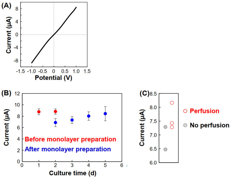
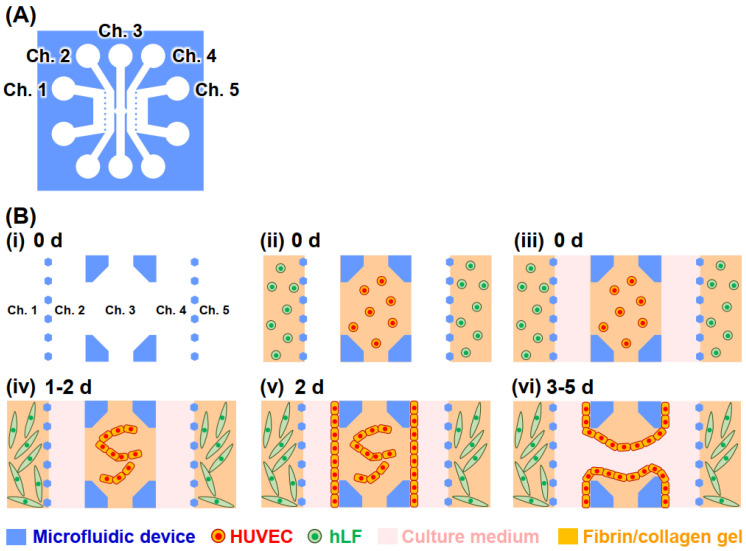
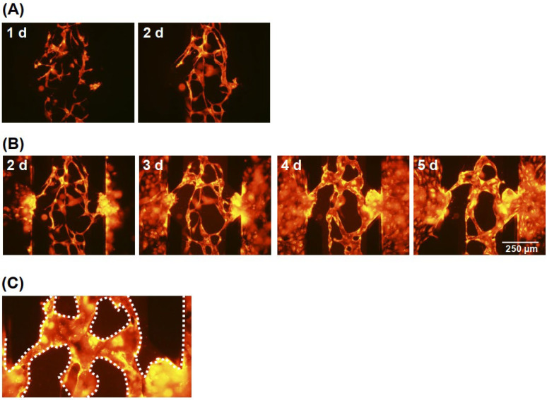
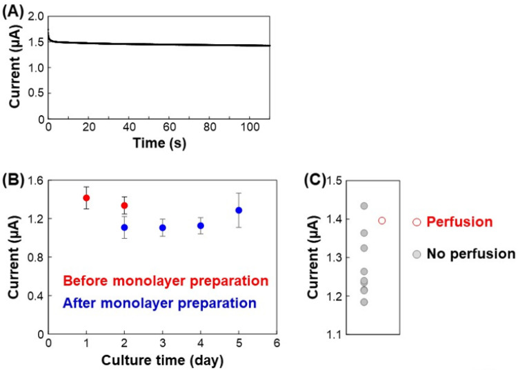
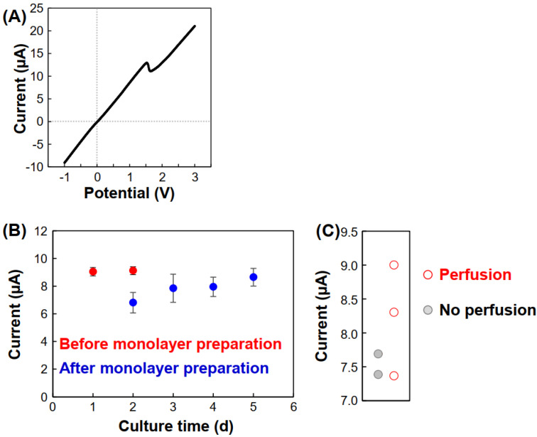
We present a novel methodology based on ion conductance to evaluate the perfusability of vascular vessels in microfluidic devices without microscopic imaging. The devices consisted of five channels, with the center channel filled with fibrin/collagen gel containing human umbilical vein endothelial cells (HUVECs). Fibroblasts were cultured in the other channels to improve the vascular network formation. To form vessel structures bridging the center channel, HUVEC monolayers were prepared on both side walls of the gel. During the culture, the HUVECs migrated from the monolayer and connected to the HUVECs in the gel, and vascular vessels formed, resulting in successful perfusion between the channels after culturing for 3-5 d. To evaluate perfusion without microscopic imaging, Ag/AgCl wires were inserted into the channels, and ion currents were obtained to measure the ion conductance between the channels separated by the HUVEC monolayers. As the HUVEC monolayers blocked the ion current flow, the ion currents were low before vessel formation. In contrast, ion currents increased after vessel formation because of creation of ion current paths. Thus, the observed ion currents were correlated with the perfusability of the vessels, indicating that they can be used as indicators of perfusion during vessel formation in microfluidic devices. The developed methodology will be used for drug screening using organs-on-a-chip containing vascular vessels.
我们提出了一种基于离子电导的新方法,用于在无微镜成像的情况下评估微流控装置中血管的灌注能力。该装置由五个通道组成,中间通道填充有含有人类脐静脉内皮细胞(HUVECs)的纤维蛋白/胶原蛋白凝胶。在其他通道中培养成纤维细胞以促进血管网络的形成。为了形成连接中间通道的血管结构,在凝胶的两侧壁上制备了HUVEC单层。在培养过程中,HUVECs从单层迁移并与凝胶中的HUVECs连接,形成血管,培养3-5天后通道之间实现了成功灌注。为了在无微镜成像的情况下评估灌注,将Ag/AgCl电极插入通道中,并获取离子电流以测量由HUVEC单层分隔的通道之间的离子电导。由于HUVEC单层阻断了离子电流,在血管形成之前离子电流较低。相反,血管形成后离子电流增加,因为形成了离子电流路径。因此,观察到的离子电流与血管的灌注能力相关,表明它们可作为微流控装置中血管形成过程中灌注的指标。所开发的方法将用于使用含有血管的芯片器官进行药物筛选。