Institute of Anatomy and Cell Biology, Julius-Maximilians-University Wuerzburg, 97070 Wuerzburg, Germany.
Eye Center, Medical Center, Faculty of Medicine, University of Freiburg, 79106 Freiburg, Germany.
Int J Mol Sci. 2021 Dec 10;22(24):13318. doi: 10.3390/ijms222413318.
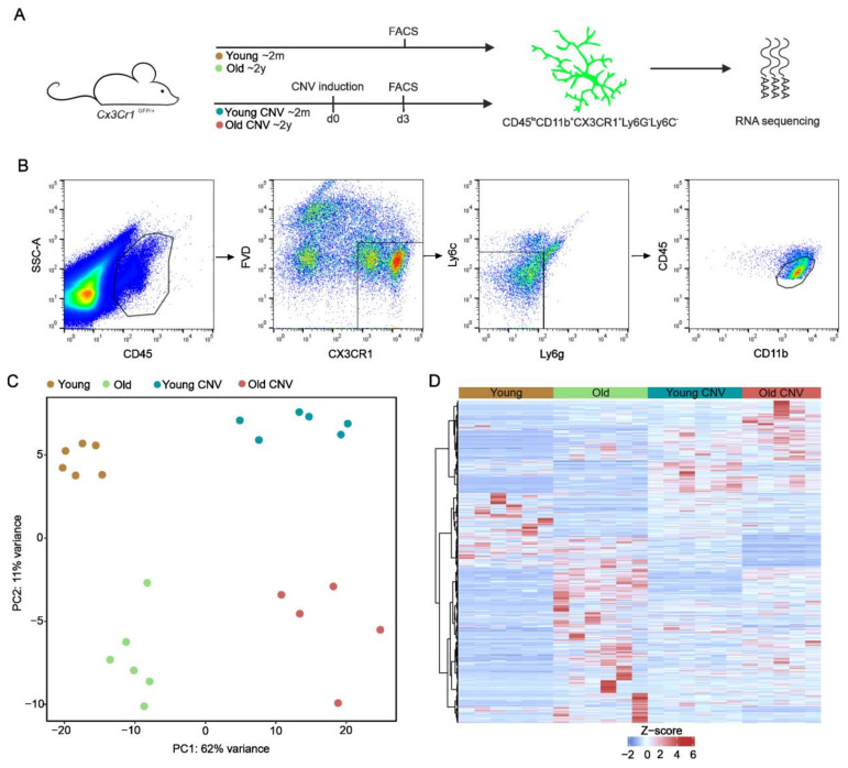
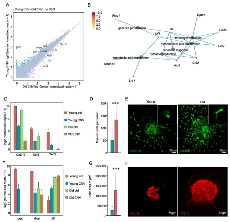
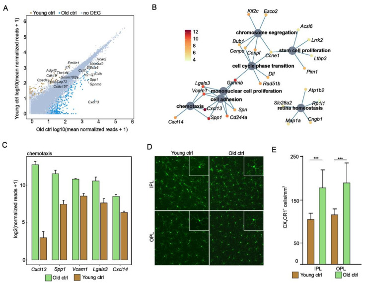
Immunosenescence is considered a possible factor in the development of age-related macular degeneration and choroidal neovascularization (CNV). However, age-related changes of myeloid cells (MCs), such as microglia and macrophages, in the healthy retina or during CNV formation are ill-defined. In this study, -positive MCs were isolated by fluorescence-activated cell sorting from six-week (young) and two-year-old (old) mice, both during physiological aging and laser-induced CNV development. High-throughput RNA-sequencing was performed to define the age-dependent transcriptional differences in MCs during physiological aging and CNV development, complemented by immunohistochemical characterization and the quantification of MCs, as well as CNV size measurements. These analyses revealed that myeloid cells change their transcriptional profile during both aging and CNV development. In the steady state, senescent MCs demonstrated an upregulation of factors contributing to cell proliferation and chemotaxis, such as , as well as the downregulation of microglial signature genes. During CNV formation, aged myeloid cells revealed a significant upregulation of angiogenic factors such as and concomitant with significantly enlarged CNV and an increased accumulation of MCs in aged mice in comparison to young mice. Future studies need to clarify whether this observation is an epiphenomenon or a causal relationship to determine the role of immunosenescence in CNV formation.
免疫衰老被认为是年龄相关性黄斑变性和脉络膜新生血管(CNV)发展的一个可能因素。然而,健康视网膜或 CNV 形成过程中髓样细胞(MCs)的年龄相关变化(如小胶质细胞和巨噬细胞)仍不明确。在这项研究中,我们通过荧光激活细胞分选从小鼠中分离出阳性 MCs,这些小鼠分别处于 6 周龄(年轻)和 2 岁(年老),同时处于生理衰老和激光诱导的 CNV 发展过程中。我们进行了高通量 RNA 测序,以确定生理衰老和 CNV 发展过程中 MCs 的年龄依赖性转录差异,并用免疫组织化学特征描述和 MCs 以及 CNV 大小的定量测量来补充这些分析。这些分析表明,髓样细胞在衰老和 CNV 发展过程中改变了其转录谱。在稳定状态下,衰老的 MCs 表现出促进细胞增殖和趋化的因子上调,如 ,以及小胶质细胞特征基因的下调。在 CNV 形成过程中,与年轻小鼠相比,衰老的髓样细胞显著上调了血管生成因子,如 和 ,同时 CNV 显著增大,MCs 在衰老小鼠中的积累增加。未来的研究需要阐明这种观察是一种偶然现象还是与确定免疫衰老在 CNV 形成中的作用有关的因果关系。